Zinc »
PDB 7dvd-7ea5 »
7e63 »
Zinc in PDB 7e63: The Crystal Structure of Peptidoglycan Peptidase in Complex with Inhibitor 2-1
Protein crystallography data
The structure of The Crystal Structure of Peptidoglycan Peptidase in Complex with Inhibitor 2-1, PDB code: 7e63
was solved by
Y.Choi,
K.J.Min,
H.J.Yoon,
H.H.Lee,
with X-Ray Crystallography technique. A brief refinement statistics is given in the table below:
| Resolution Low / High (Å) | 67.48 / 2.40 |
| Space group | P 21 21 21 |
| Cell size a, b, c (Å), α, β, γ (°) | 56.872, 90.734, 100.946, 90, 90, 90 |
| R / Rfree (%) | 19.3 / 26.5 |
Zinc Binding Sites:
The binding sites of Zinc atom in the The Crystal Structure of Peptidoglycan Peptidase in Complex with Inhibitor 2-1
(pdb code 7e63). This binding sites where shown within
5.0 Angstroms radius around Zinc atom.
In total 2 binding sites of Zinc where determined in the The Crystal Structure of Peptidoglycan Peptidase in Complex with Inhibitor 2-1, PDB code: 7e63:
Jump to Zinc binding site number: 1; 2;
In total 2 binding sites of Zinc where determined in the The Crystal Structure of Peptidoglycan Peptidase in Complex with Inhibitor 2-1, PDB code: 7e63:
Jump to Zinc binding site number: 1; 2;
Zinc binding site 1 out of 2 in 7e63
Go back to
Zinc binding site 1 out
of 2 in the The Crystal Structure of Peptidoglycan Peptidase in Complex with Inhibitor 2-1
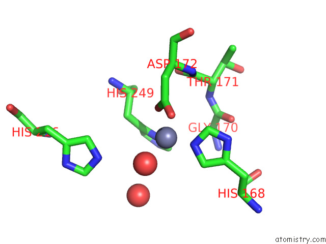
Mono view
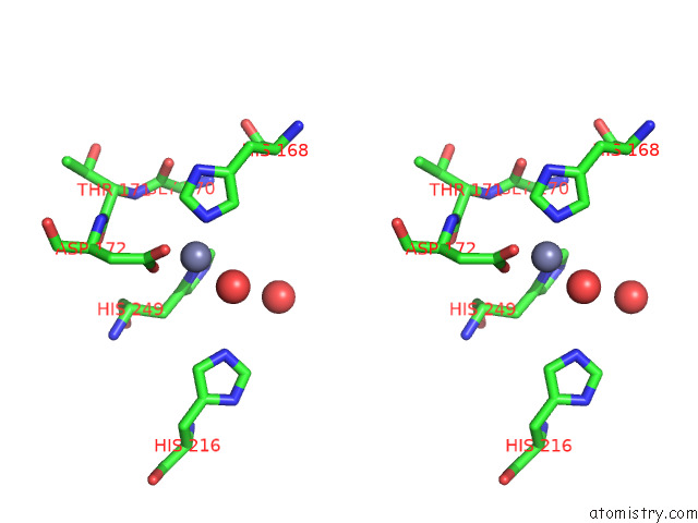
Stereo pair view
Mono view
Stereo pair view
A full contact list of Zinc with other atoms in the Zn binding
site number 1 of The Crystal Structure of Peptidoglycan Peptidase in Complex with Inhibitor 2-1 within 5.0Å range:
|
Zinc binding site 2 out of 2 in 7e63
Go back to
Zinc binding site 2 out
of 2 in the The Crystal Structure of Peptidoglycan Peptidase in Complex with Inhibitor 2-1
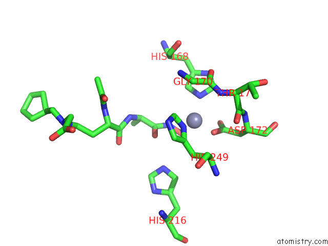
Mono view
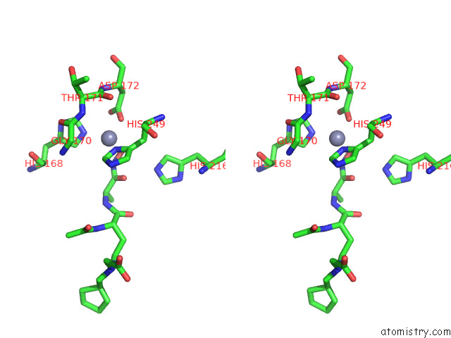
Stereo pair view
Mono view
Stereo pair view
A full contact list of Zinc with other atoms in the Zn binding
site number 2 of The Crystal Structure of Peptidoglycan Peptidase in Complex with Inhibitor 2-1 within 5.0Å range:
|
Reference:
Y.Choi,
J.S.Park,
J.Kim,
K.Min,
K.Mahasenan,
C.Kim,
H.J.Yoon,
S.Lim,
D.H.Cheon,
Y.Lee,
S.Ryu,
S.Mobashery,
B.M.Kim,
H.H.Lee.
Structure-Based Inhibitor Design For Reshaping Bacterial Morphology Commun Biol V. 5 395 2022.
ISSN: ESSN 2399-3642
DOI: 10.1038/S42003-022-03355-3
Page generated: Tue Oct 29 19:36:06 2024
ISSN: ESSN 2399-3642
DOI: 10.1038/S42003-022-03355-3
Last articles
Zn in 9JYWZn in 9IR4
Zn in 9IR3
Zn in 9GMX
Zn in 9GMW
Zn in 9JEJ
Zn in 9ERF
Zn in 9ERE
Zn in 9EGV
Zn in 9EGW