Zinc »
PDB 7dvd-7ea5 »
7e3u »
Zinc in PDB 7e3u: Crystal Structure of the Pseudomonas Aeruginosa Dihydropyrimidinase Complexed with 5-Au
Enzymatic activity of Crystal Structure of the Pseudomonas Aeruginosa Dihydropyrimidinase Complexed with 5-Au
All present enzymatic activity of Crystal Structure of the Pseudomonas Aeruginosa Dihydropyrimidinase Complexed with 5-Au:
3.5.2.2;
3.5.2.2;
Protein crystallography data
The structure of Crystal Structure of the Pseudomonas Aeruginosa Dihydropyrimidinase Complexed with 5-Au, PDB code: 7e3u
was solved by
Y.C.Yang,
R.H.Luo,
Y.H.Huang,
C.Y.Huang,
E.S.Lin,
with X-Ray Crystallography technique. A brief refinement statistics is given in the table below:
| Resolution Low / High (Å) | 28.01 / 2.16 |
| Space group | P 31 2 1 |
| Cell size a, b, c (Å), α, β, γ (°) | 112.667, 112.667, 161.432, 90, 90, 120 |
| R / Rfree (%) | 18.6 / 23 |
Zinc Binding Sites:
The binding sites of Zinc atom in the Crystal Structure of the Pseudomonas Aeruginosa Dihydropyrimidinase Complexed with 5-Au
(pdb code 7e3u). This binding sites where shown within
5.0 Angstroms radius around Zinc atom.
In total 4 binding sites of Zinc where determined in the Crystal Structure of the Pseudomonas Aeruginosa Dihydropyrimidinase Complexed with 5-Au, PDB code: 7e3u:
Jump to Zinc binding site number: 1; 2; 3; 4;
In total 4 binding sites of Zinc where determined in the Crystal Structure of the Pseudomonas Aeruginosa Dihydropyrimidinase Complexed with 5-Au, PDB code: 7e3u:
Jump to Zinc binding site number: 1; 2; 3; 4;
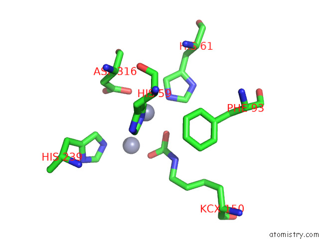
Zinc binding site 1 out of 4 in 7e3u
Go back to
Zinc binding site 1 out
of 4 in the Crystal Structure of the Pseudomonas Aeruginosa Dihydropyrimidinase Complexed with 5-Au
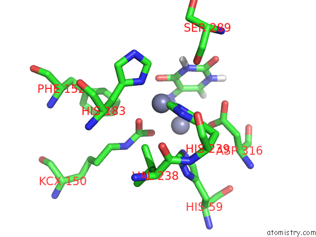
Mono view
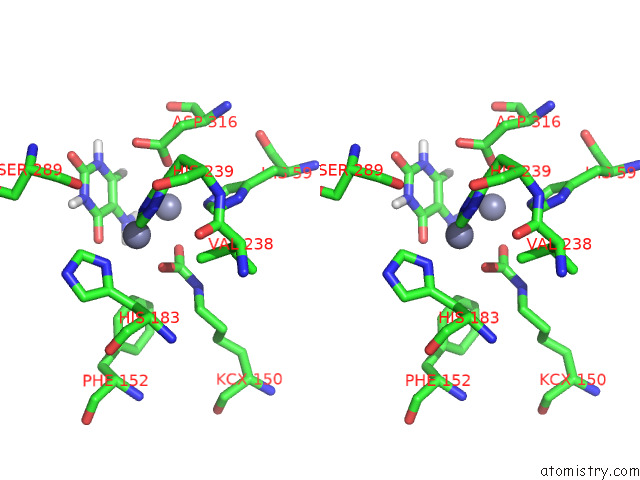
Stereo pair view
Mono view
Stereo pair view
A full contact list of Zinc with other atoms in the Zn binding
site number 1 of Crystal Structure of the Pseudomonas Aeruginosa Dihydropyrimidinase Complexed with 5-Au within 5.0Å range:
|
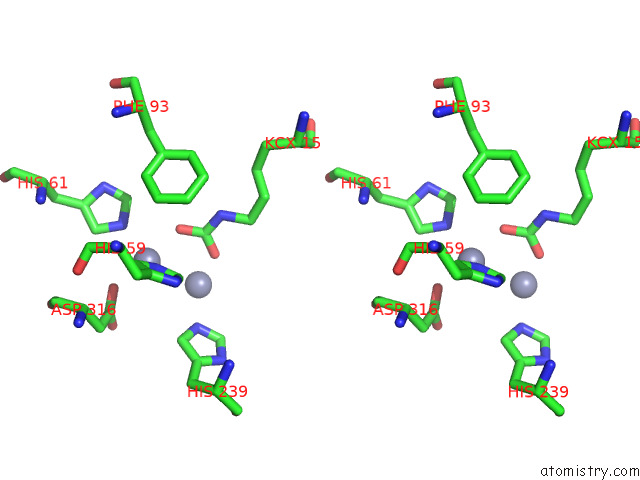
Zinc binding site 2 out of 4 in 7e3u
Go back to
Zinc binding site 2 out
of 4 in the Crystal Structure of the Pseudomonas Aeruginosa Dihydropyrimidinase Complexed with 5-Au
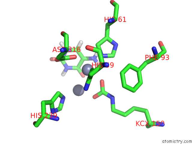
Mono view
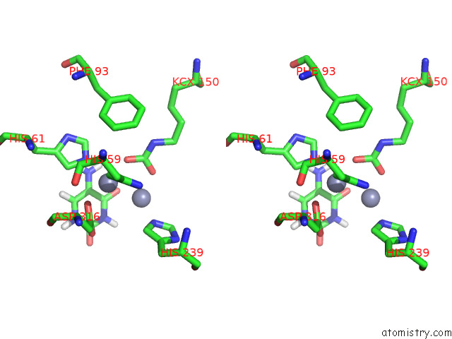
Stereo pair view
Mono view
Stereo pair view
A full contact list of Zinc with other atoms in the Zn binding
site number 2 of Crystal Structure of the Pseudomonas Aeruginosa Dihydropyrimidinase Complexed with 5-Au within 5.0Å range:
|
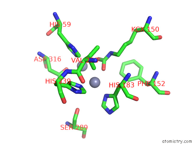
Zinc binding site 3 out of 4 in 7e3u
Go back to
Zinc binding site 3 out
of 4 in the Crystal Structure of the Pseudomonas Aeruginosa Dihydropyrimidinase Complexed with 5-Au
Mono view
Stereo pair view
Mono view
Stereo pair view
A full contact list of Zinc with other atoms in the Zn binding
site number 3 of Crystal Structure of the Pseudomonas Aeruginosa Dihydropyrimidinase Complexed with 5-Au within 5.0Å range:
|
Zinc binding site 4 out of 4 in 7e3u
Go back to
Zinc binding site 4 out
of 4 in the Crystal Structure of the Pseudomonas Aeruginosa Dihydropyrimidinase Complexed with 5-Au
Mono view
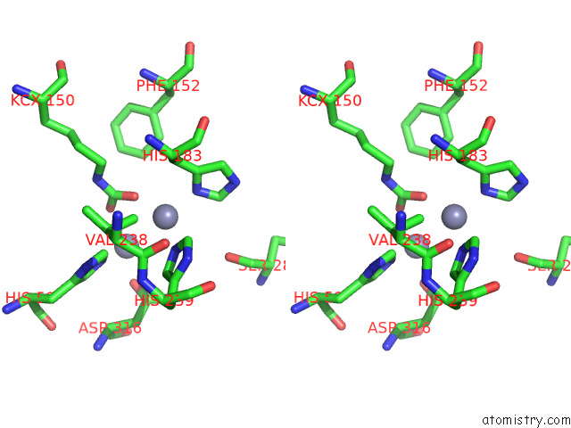
Stereo pair view
Mono view
Stereo pair view
A full contact list of Zinc with other atoms in the Zn binding
site number 4 of Crystal Structure of the Pseudomonas Aeruginosa Dihydropyrimidinase Complexed with 5-Au within 5.0Å range:
|
Reference:
E.S.Lin,
R.H.Luo,
Y.C.Yang,
C.Y.Huang.
Molecular Insights Into How the Dimetal Center in Dihydropyrimidinase Can Bind the Thymine Antagonist 5-Aminouracil: A Different Binding Mode From the Anticancer Drug 5-Fluorouracil. Bioinorg Chem Appl V.2022 17745 2022.
ISSN: ISSN 1565-3633
PubMed: 35198016
DOI: 10.1155/2022/1817745
Page generated: Tue Oct 29 19:33:05 2024
ISSN: ISSN 1565-3633
PubMed: 35198016
DOI: 10.1155/2022/1817745
Last articles
Zn in 9JYWZn in 9IR4
Zn in 9IR3
Zn in 9GMX
Zn in 9GMW
Zn in 9JEJ
Zn in 9ERF
Zn in 9ERE
Zn in 9EGV
Zn in 9EGW