Zinc »
PDB 2b65-2bmi »
2bh7 »
Zinc in PDB 2bh7: Crystal Structure of A Semet Derivative of Amid at 2.2 Angstroms
Enzymatic activity of Crystal Structure of A Semet Derivative of Amid at 2.2 Angstroms
All present enzymatic activity of Crystal Structure of A Semet Derivative of Amid at 2.2 Angstroms:
3.5.1.28;
3.5.1.28;
Protein crystallography data
The structure of Crystal Structure of A Semet Derivative of Amid at 2.2 Angstroms, PDB code: 2bh7
was solved by
S.Petrella,
R.Herman,
E.Sauvage,
C.Genereux,
A.Pennartz,
B.Joris,
P.Charlier,
with X-Ray Crystallography technique. A brief refinement statistics is given in the table below:
| Resolution Low / High (Å) | 27.77 / 2.20 |
| Space group | P 61 2 2 |
| Cell size a, b, c (Å), α, β, γ (°) | 88.990, 88.990, 183.923, 90.00, 90.00, 120.00 |
| R / Rfree (%) | 23.1 / 27.9 |
Zinc Binding Sites:
The binding sites of Zinc atom in the Crystal Structure of A Semet Derivative of Amid at 2.2 Angstroms
(pdb code 2bh7). This binding sites where shown within
5.0 Angstroms radius around Zinc atom.
In total only one binding site of Zinc was determined in the Crystal Structure of A Semet Derivative of Amid at 2.2 Angstroms, PDB code: 2bh7:
In total only one binding site of Zinc was determined in the Crystal Structure of A Semet Derivative of Amid at 2.2 Angstroms, PDB code: 2bh7:
Zinc binding site 1 out of 1 in 2bh7
Go back to
Zinc binding site 1 out
of 1 in the Crystal Structure of A Semet Derivative of Amid at 2.2 Angstroms
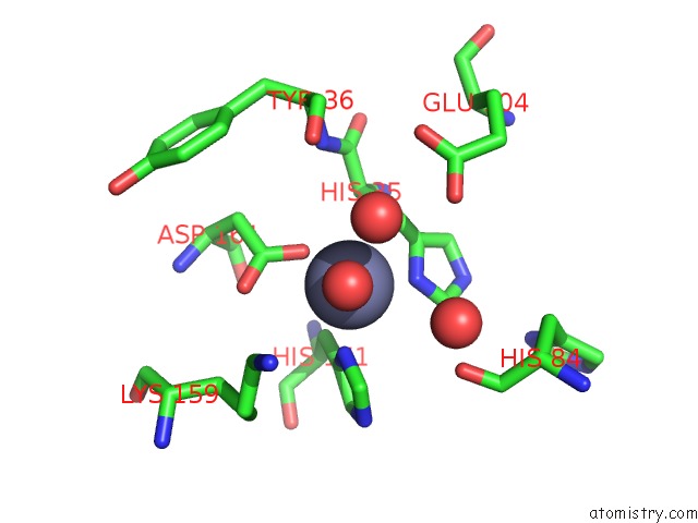
Mono view
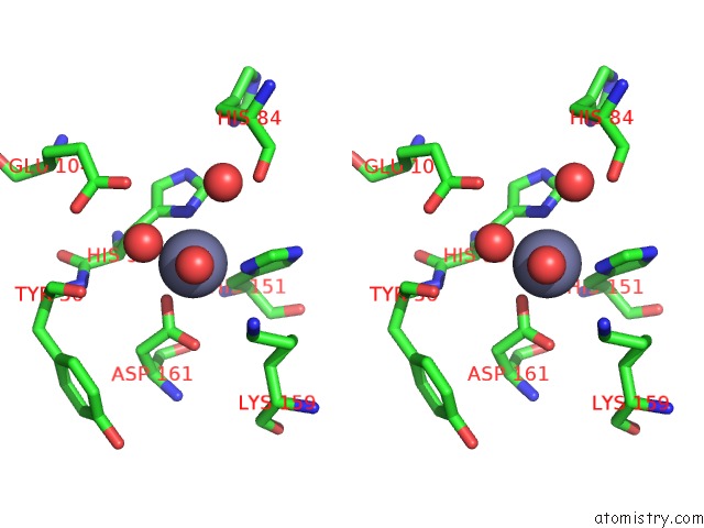
Stereo pair view
Mono view
Stereo pair view
A full contact list of Zinc with other atoms in the Zn binding
site number 1 of Crystal Structure of A Semet Derivative of Amid at 2.2 Angstroms within 5.0Å range:
|
Reference:
F.Kerff,
S.Petrella,
F.Mercier,
E.Sauvage,
R.Herman,
A.Pennartz,
A.Zervosen,
A.Luxen,
J.M.Frere,
B.Joris,
P.Charlier.
Specific Structural Features of the N-Acetylmuramoyl-L-Alanine Amidase Amid From Escherichia Coli and Mechanistic Implications For Enzymes of This Family. J.Mol.Biol. V. 397 249 2010.
ISSN: ISSN 0022-2836
PubMed: 20036252
DOI: 10.1016/J.JMB.2009.12.038
Page generated: Wed Oct 16 22:01:51 2024
ISSN: ISSN 0022-2836
PubMed: 20036252
DOI: 10.1016/J.JMB.2009.12.038
Last articles
Zn in 9JYWZn in 9IR4
Zn in 9IR3
Zn in 9GMX
Zn in 9GMW
Zn in 9JEJ
Zn in 9ERF
Zn in 9ERE
Zn in 9EGV
Zn in 9EGW