Zinc »
PDB 2b65-2bmi »
2b9d »
Zinc in PDB 2b9d: Crystal Structure of Hpv E7 CR3 Domain
Protein crystallography data
The structure of Crystal Structure of Hpv E7 CR3 Domain, PDB code: 2b9d
was solved by
X.Liu,
A.Clements,
K.Zhao,
R.Marmorstein,
with X-Ray Crystallography technique. A brief refinement statistics is given in the table below:
| Resolution Low / High (Å) | 50.00 / 1.60 |
| Space group | P 21 21 2 |
| Cell size a, b, c (Å), α, β, γ (°) | 33.908, 44.935, 58.670, 90.00, 90.00, 90.00 |
| R / Rfree (%) | 20.8 / 25.2 |
Zinc Binding Sites:
The binding sites of Zinc atom in the Crystal Structure of Hpv E7 CR3 Domain
(pdb code 2b9d). This binding sites where shown within
5.0 Angstroms radius around Zinc atom.
In total 2 binding sites of Zinc where determined in the Crystal Structure of Hpv E7 CR3 Domain, PDB code: 2b9d:
Jump to Zinc binding site number: 1; 2;
In total 2 binding sites of Zinc where determined in the Crystal Structure of Hpv E7 CR3 Domain, PDB code: 2b9d:
Jump to Zinc binding site number: 1; 2;
Zinc binding site 1 out of 2 in 2b9d
Go back to
Zinc binding site 1 out
of 2 in the Crystal Structure of Hpv E7 CR3 Domain
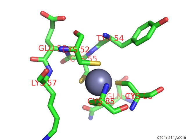
Mono view
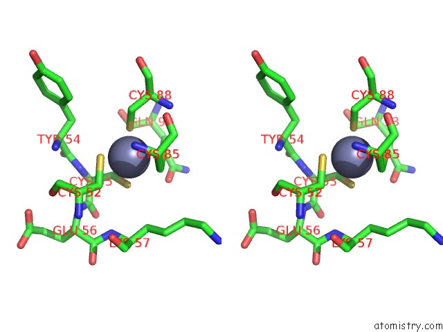
Stereo pair view
Mono view
Stereo pair view
A full contact list of Zinc with other atoms in the Zn binding
site number 1 of Crystal Structure of Hpv E7 CR3 Domain within 5.0Å range:
|
Zinc binding site 2 out of 2 in 2b9d
Go back to
Zinc binding site 2 out
of 2 in the Crystal Structure of Hpv E7 CR3 Domain

Mono view

Stereo pair view
Mono view
Stereo pair view
A full contact list of Zinc with other atoms in the Zn binding
site number 2 of Crystal Structure of Hpv E7 CR3 Domain within 5.0Å range:
|
Reference:
X.Liu,
A.Clements,
K.Zhao,
R.Marmorstein.
Structure of the Human Papillomavirus E7 Oncoprotein and Its Mechanism For Inactivation of the Retinoblastoma Tumor Suppressor. J.Biol.Chem. V. 281 578 2006.
ISSN: ISSN 0021-9258
PubMed: 16249186
DOI: 10.1074/JBC.M508455200
Page generated: Wed Oct 16 21:56:47 2024
ISSN: ISSN 0021-9258
PubMed: 16249186
DOI: 10.1074/JBC.M508455200
Last articles
Zn in 9JYWZn in 9IR4
Zn in 9IR3
Zn in 9GMX
Zn in 9GMW
Zn in 9JEJ
Zn in 9ERF
Zn in 9ERE
Zn in 9EGV
Zn in 9EGW