Zinc »
PDB 7y81-7yj3 »
7ygi »
Zinc in PDB 7ygi: Crystal Structure of P53 Dbd Domain in Complex with Azurin
Protein crystallography data
The structure of Crystal Structure of P53 Dbd Domain in Complex with Azurin, PDB code: 7ygi
was solved by
W.X.Jiang,
J.Q.Zuo,
J.J.Hu,
X.Q.Chen,
L.X.Ma,
Z.Liu,
Q.Xing,
with X-Ray Crystallography technique. A brief refinement statistics is given in the table below:
| Resolution Low / High (Å) | 19.88 / 2.10 |
| Space group | C 1 2 1 |
| Cell size a, b, c (Å), α, β, γ (°) | 144.839, 68.75, 83.847, 90, 99.69, 90 |
| R / Rfree (%) | 21.3 / 27 |
Other elements in 7ygi:
The structure of Crystal Structure of P53 Dbd Domain in Complex with Azurin also contains other interesting chemical elements:
| Sodium | (Na) | 2 atoms |
| Potassium | (K) | 2 atoms |
Zinc Binding Sites:
The binding sites of Zinc atom in the Crystal Structure of P53 Dbd Domain in Complex with Azurin
(pdb code 7ygi). This binding sites where shown within
5.0 Angstroms radius around Zinc atom.
In total 2 binding sites of Zinc where determined in the Crystal Structure of P53 Dbd Domain in Complex with Azurin, PDB code: 7ygi:
Jump to Zinc binding site number: 1; 2;
In total 2 binding sites of Zinc where determined in the Crystal Structure of P53 Dbd Domain in Complex with Azurin, PDB code: 7ygi:
Jump to Zinc binding site number: 1; 2;
Zinc binding site 1 out of 2 in 7ygi
Go back to
Zinc binding site 1 out
of 2 in the Crystal Structure of P53 Dbd Domain in Complex with Azurin
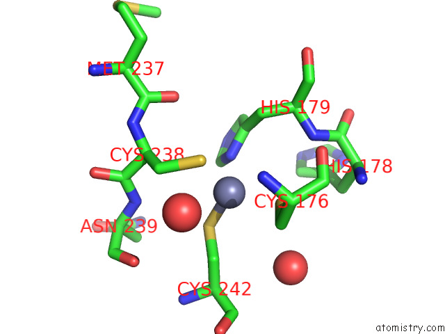
Mono view
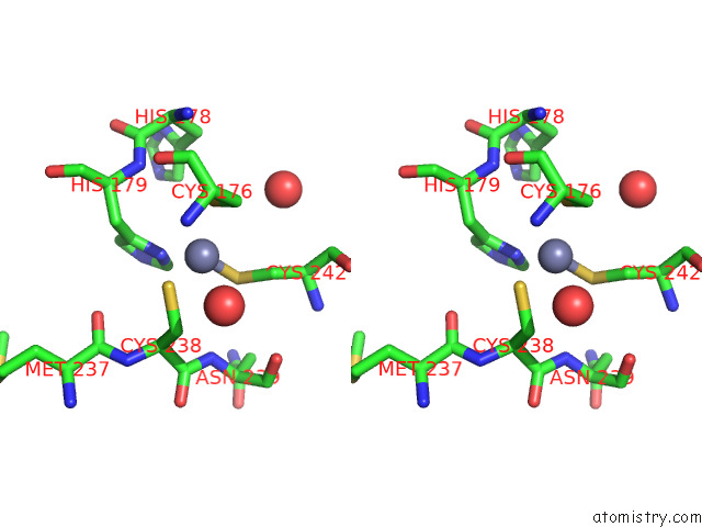
Stereo pair view
Mono view
Stereo pair view
A full contact list of Zinc with other atoms in the Zn binding
site number 1 of Crystal Structure of P53 Dbd Domain in Complex with Azurin within 5.0Å range:
|
Zinc binding site 2 out of 2 in 7ygi
Go back to
Zinc binding site 2 out
of 2 in the Crystal Structure of P53 Dbd Domain in Complex with Azurin
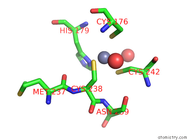
Mono view
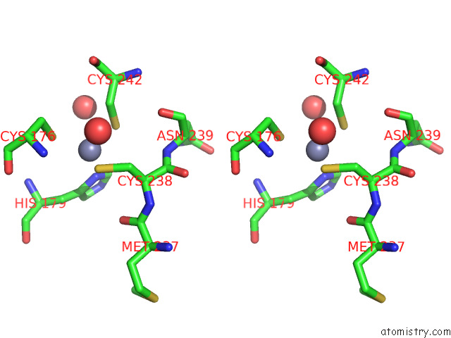
Stereo pair view
Mono view
Stereo pair view
A full contact list of Zinc with other atoms in the Zn binding
site number 2 of Crystal Structure of P53 Dbd Domain in Complex with Azurin within 5.0Å range:
|
Reference:
J.Hu,
W.Jiang,
J.Zuo,
D.Shi,
X.Chen,
X.Yang,
W.Zhang,
L.Ma,
Z.Liu,
Q.Xing.
Structural Basis of Bacterial Effector Protein Azurin Targeting Tumor Suppressor P53 and Inhibiting Its Ubiquitination. Commun Biol V. 6 59 2023.
ISSN: ESSN 2399-3642
PubMed: 36650277
DOI: 10.1038/S42003-023-04458-1
Page generated: Wed Oct 30 15:49:09 2024
ISSN: ESSN 2399-3642
PubMed: 36650277
DOI: 10.1038/S42003-023-04458-1
Last articles
Zn in 9JYWZn in 9IR4
Zn in 9IR3
Zn in 9GMX
Zn in 9GMW
Zn in 9JEJ
Zn in 9ERF
Zn in 9ERE
Zn in 9EGV
Zn in 9EGW