Zinc »
PDB 7wm7-7wun »
7wt5 »
Zinc in PDB 7wt5: Crystal Structure of Hla-A*2450 Complexed with 8-Mer Model Peptide
Protein crystallography data
The structure of Crystal Structure of Hla-A*2450 Complexed with 8-Mer Model Peptide, PDB code: 7wt5
was solved by
M.Asa,
D.Morita,
M.Sugita,
with X-Ray Crystallography technique. A brief refinement statistics is given in the table below:
| Resolution Low / High (Å) | 45.81 / 2.10 |
| Space group | P 1 21 1 |
| Cell size a, b, c (Å), α, β, γ (°) | 86.239, 46.46, 141.65, 90, 104.01, 90 |
| R / Rfree (%) | 18.6 / 22.8 |
Zinc Binding Sites:
The binding sites of Zinc atom in the Crystal Structure of Hla-A*2450 Complexed with 8-Mer Model Peptide
(pdb code 7wt5). This binding sites where shown within
5.0 Angstroms radius around Zinc atom.
In total 3 binding sites of Zinc where determined in the Crystal Structure of Hla-A*2450 Complexed with 8-Mer Model Peptide, PDB code: 7wt5:
Jump to Zinc binding site number: 1; 2; 3;
In total 3 binding sites of Zinc where determined in the Crystal Structure of Hla-A*2450 Complexed with 8-Mer Model Peptide, PDB code: 7wt5:
Jump to Zinc binding site number: 1; 2; 3;
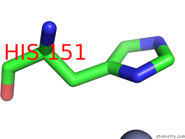
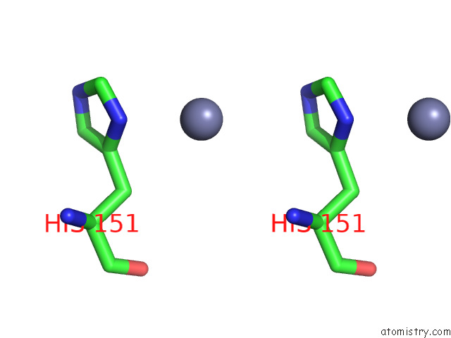
Zinc binding site 1 out of 3 in 7wt5
Go back to
Zinc binding site 1 out
of 3 in the Crystal Structure of Hla-A*2450 Complexed with 8-Mer Model Peptide
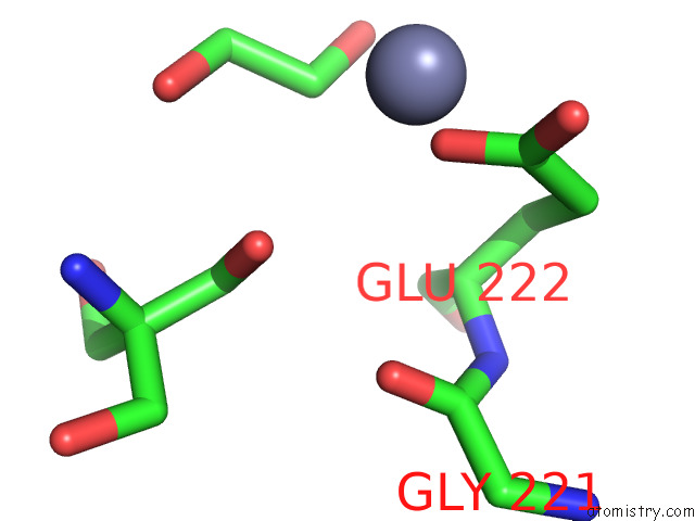
Mono view
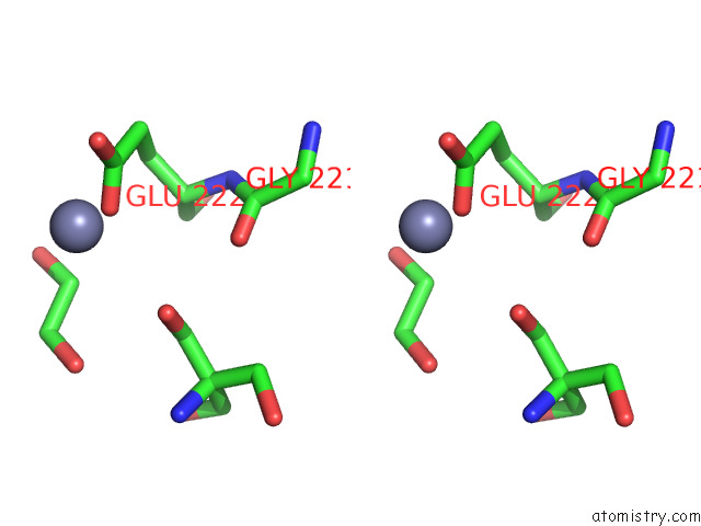
Stereo pair view
Mono view
Stereo pair view
A full contact list of Zinc with other atoms in the Zn binding
site number 1 of Crystal Structure of Hla-A*2450 Complexed with 8-Mer Model Peptide within 5.0Å range:
|
Zinc binding site 2 out of 3 in 7wt5
Go back to
Zinc binding site 2 out
of 3 in the Crystal Structure of Hla-A*2450 Complexed with 8-Mer Model Peptide
Mono view
Stereo pair view
Mono view
Stereo pair view
A full contact list of Zinc with other atoms in the Zn binding
site number 2 of Crystal Structure of Hla-A*2450 Complexed with 8-Mer Model Peptide within 5.0Å range:
|
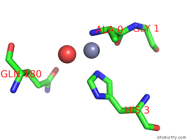
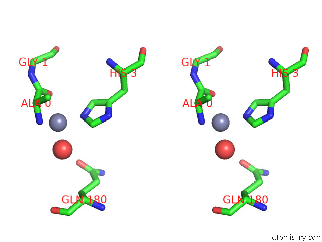
Zinc binding site 3 out of 3 in 7wt5
Go back to
Zinc binding site 3 out
of 3 in the Crystal Structure of Hla-A*2450 Complexed with 8-Mer Model Peptide
Mono view
Stereo pair view
Mono view
Stereo pair view
A full contact list of Zinc with other atoms in the Zn binding
site number 3 of Crystal Structure of Hla-A*2450 Complexed with 8-Mer Model Peptide within 5.0Å range:
|
Reference:
M.Asa,
D.Morita,
J.Kuroha,
T.Mizutani,
N.Mori,
B.Mikami,
M.Sugita.
Crystal Structures of N-Myristoylated Lipopeptide-Bound Hla Class I Complexes Indicate Reorganization of B-Pocket Architecture Upon Ligand Binding. J.Biol.Chem. V. 298 02100 2022.
ISSN: ESSN 1083-351X
PubMed: 35667438
DOI: 10.1016/J.JBC.2022.102100
Page generated: Wed Oct 30 14:35:30 2024
ISSN: ESSN 1083-351X
PubMed: 35667438
DOI: 10.1016/J.JBC.2022.102100
Last articles
Zn in 9JYWZn in 9IR4
Zn in 9IR3
Zn in 9GMX
Zn in 9GMW
Zn in 9JEJ
Zn in 9ERF
Zn in 9ERE
Zn in 9EGV
Zn in 9EGW