Zinc »
PDB 7t08-7tln »
7t9x »
Zinc in PDB 7t9x: Saccharomyces Cerevisiae PEX12 Ring Domain
Protein crystallography data
The structure of Saccharomyces Cerevisiae PEX12 Ring Domain, PDB code: 7t9x
was solved by
P.Feng,
T.Rapoport,
with X-Ray Crystallography technique. A brief refinement statistics is given in the table below:
| Resolution Low / High (Å) | 71.22 / 1.52 |
| Space group | P 1 21 1 |
| Cell size a, b, c (Å), α, β, γ (°) | 27.578, 32.922, 72.488, 90, 100.72, 90 |
| R / Rfree (%) | 18.6 / 19.7 |
Zinc Binding Sites:
The binding sites of Zinc atom in the Saccharomyces Cerevisiae PEX12 Ring Domain
(pdb code 7t9x). This binding sites where shown within
5.0 Angstroms radius around Zinc atom.
In total 2 binding sites of Zinc where determined in the Saccharomyces Cerevisiae PEX12 Ring Domain, PDB code: 7t9x:
Jump to Zinc binding site number: 1; 2;
In total 2 binding sites of Zinc where determined in the Saccharomyces Cerevisiae PEX12 Ring Domain, PDB code: 7t9x:
Jump to Zinc binding site number: 1; 2;
Zinc binding site 1 out of 2 in 7t9x
Go back to
Zinc binding site 1 out
of 2 in the Saccharomyces Cerevisiae PEX12 Ring Domain
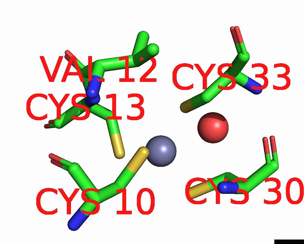
Mono view
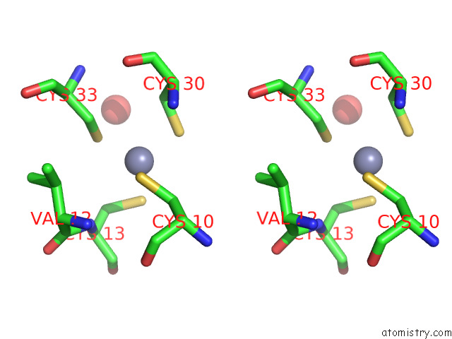
Stereo pair view
Mono view
Stereo pair view
A full contact list of Zinc with other atoms in the Zn binding
site number 1 of Saccharomyces Cerevisiae PEX12 Ring Domain within 5.0Å range:
|
Zinc binding site 2 out of 2 in 7t9x
Go back to
Zinc binding site 2 out
of 2 in the Saccharomyces Cerevisiae PEX12 Ring Domain
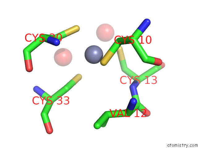
Mono view
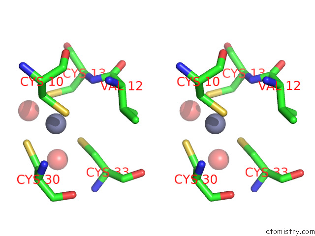
Stereo pair view
Mono view
Stereo pair view
A full contact list of Zinc with other atoms in the Zn binding
site number 2 of Saccharomyces Cerevisiae PEX12 Ring Domain within 5.0Å range:
|
Reference:
P.Feng,
X.Wu,
S.K.Erramilli,
J.A.Paulo,
P.Knejski,
S.P.Gygi,
A.A.Kossiakoff,
T.A.Rapoport.
A Peroxisomal Ubiquitin Ligase Complex Forms A Retrotranslocation Channel. Nature V. 607 374 2022.
ISSN: ESSN 1476-4687
PubMed: 35768507
DOI: 10.1038/S41586-022-04903-X
Page generated: Wed Oct 30 11:33:00 2024
ISSN: ESSN 1476-4687
PubMed: 35768507
DOI: 10.1038/S41586-022-04903-X
Last articles
Zn in 9JYWZn in 9IR4
Zn in 9IR3
Zn in 9GMX
Zn in 9GMW
Zn in 9JEJ
Zn in 9ERF
Zn in 9ERE
Zn in 9EGV
Zn in 9EGW