Zinc »
PDB 7khc-7kuq »
7knf »
Zinc in PDB 7knf: 1.80A Resolution Structure of Independent Phosphoglycerate Mutase From C. Elegans in Complex with A Macrocyclic Peptide Inhibitor (Ce-1 Nhoh)
Enzymatic activity of 1.80A Resolution Structure of Independent Phosphoglycerate Mutase From C. Elegans in Complex with A Macrocyclic Peptide Inhibitor (Ce-1 Nhoh)
All present enzymatic activity of 1.80A Resolution Structure of Independent Phosphoglycerate Mutase From C. Elegans in Complex with A Macrocyclic Peptide Inhibitor (Ce-1 Nhoh):
5.4.2.12;
5.4.2.12;
Protein crystallography data
The structure of 1.80A Resolution Structure of Independent Phosphoglycerate Mutase From C. Elegans in Complex with A Macrocyclic Peptide Inhibitor (Ce-1 Nhoh), PDB code: 7knf
was solved by
S.Lovell,
M.M.Kashipathy,
K.P.Battaile,
M.Weidmann,
P.Dranchak,
M.Aitha,
B.Queme,
C.D.Collmus,
L.Kanter,
L.Lamy,
D.Tao,
G.Rai,
H.Suga,
J.Inglese,
with X-Ray Crystallography technique. A brief refinement statistics is given in the table below:
| Resolution Low / High (Å) | 44.13 / 1.80 |
| Space group | P 1 2 1 |
| Cell size a, b, c (Å), α, β, γ (°) | 73.669, 75.846, 100.424, 90, 98.31, 90 |
| R / Rfree (%) | 15.2 / 19.4 |
Other elements in 7knf:
The structure of 1.80A Resolution Structure of Independent Phosphoglycerate Mutase From C. Elegans in Complex with A Macrocyclic Peptide Inhibitor (Ce-1 Nhoh) also contains other interesting chemical elements:
| Sodium | (Na) | 4 atoms |
Zinc Binding Sites:
The binding sites of Zinc atom in the 1.80A Resolution Structure of Independent Phosphoglycerate Mutase From C. Elegans in Complex with A Macrocyclic Peptide Inhibitor (Ce-1 Nhoh)
(pdb code 7knf). This binding sites where shown within
5.0 Angstroms radius around Zinc atom.
In total 4 binding sites of Zinc where determined in the 1.80A Resolution Structure of Independent Phosphoglycerate Mutase From C. Elegans in Complex with A Macrocyclic Peptide Inhibitor (Ce-1 Nhoh), PDB code: 7knf:
Jump to Zinc binding site number: 1; 2; 3; 4;
In total 4 binding sites of Zinc where determined in the 1.80A Resolution Structure of Independent Phosphoglycerate Mutase From C. Elegans in Complex with A Macrocyclic Peptide Inhibitor (Ce-1 Nhoh), PDB code: 7knf:
Jump to Zinc binding site number: 1; 2; 3; 4;
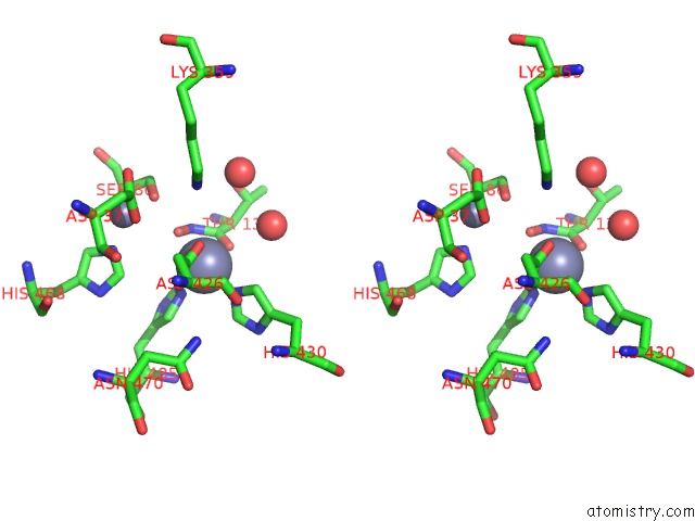
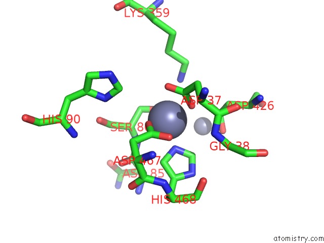
Zinc binding site 1 out of 4 in 7knf
Go back to
Zinc binding site 1 out
of 4 in the 1.80A Resolution Structure of Independent Phosphoglycerate Mutase From C. Elegans in Complex with A Macrocyclic Peptide Inhibitor (Ce-1 Nhoh)

Mono view

Stereo pair view
Mono view
Stereo pair view
A full contact list of Zinc with other atoms in the Zn binding
site number 1 of 1.80A Resolution Structure of Independent Phosphoglycerate Mutase From C. Elegans in Complex with A Macrocyclic Peptide Inhibitor (Ce-1 Nhoh) within 5.0Å range:
|
Zinc binding site 2 out of 4 in 7knf
Go back to
Zinc binding site 2 out
of 4 in the 1.80A Resolution Structure of Independent Phosphoglycerate Mutase From C. Elegans in Complex with A Macrocyclic Peptide Inhibitor (Ce-1 Nhoh)

Mono view
Stereo pair view
Mono view
Stereo pair view
A full contact list of Zinc with other atoms in the Zn binding
site number 2 of 1.80A Resolution Structure of Independent Phosphoglycerate Mutase From C. Elegans in Complex with A Macrocyclic Peptide Inhibitor (Ce-1 Nhoh) within 5.0Å range:
|
Zinc binding site 3 out of 4 in 7knf
Go back to
Zinc binding site 3 out
of 4 in the 1.80A Resolution Structure of Independent Phosphoglycerate Mutase From C. Elegans in Complex with A Macrocyclic Peptide Inhibitor (Ce-1 Nhoh)
Mono view

Stereo pair view
Mono view
Stereo pair view
A full contact list of Zinc with other atoms in the Zn binding
site number 3 of 1.80A Resolution Structure of Independent Phosphoglycerate Mutase From C. Elegans in Complex with A Macrocyclic Peptide Inhibitor (Ce-1 Nhoh) within 5.0Å range:
|
Zinc binding site 4 out of 4 in 7knf
Go back to
Zinc binding site 4 out
of 4 in the 1.80A Resolution Structure of Independent Phosphoglycerate Mutase From C. Elegans in Complex with A Macrocyclic Peptide Inhibitor (Ce-1 Nhoh)

Mono view

Stereo pair view
Mono view
Stereo pair view
A full contact list of Zinc with other atoms in the Zn binding
site number 4 of 1.80A Resolution Structure of Independent Phosphoglycerate Mutase From C. Elegans in Complex with A Macrocyclic Peptide Inhibitor (Ce-1 Nhoh) within 5.0Å range:
|
Reference:
M.Weidmann,
P.Dranchak,
M.Aitha,
B.Queme,
C.D.Collmus,
L.Kanter,
L.Lamy,
D.Tao,
M.M.Kashipathy,
K.P.Battaile,
G.Rai,
S.Lovell,
H.Suga,
J.Inglese.
Structure-Activity Relationship of Ipglycermide Binding to Phosphoglycerate Mutases J.Biol.Chem. 2021.
ISSN: ESSN 1083-351X
Page generated: Tue Oct 29 22:24:22 2024
ISSN: ESSN 1083-351X
Last articles
Zn in 9JYWZn in 9IR4
Zn in 9IR3
Zn in 9GMX
Zn in 9GMW
Zn in 9JEJ
Zn in 9ERF
Zn in 9ERE
Zn in 9EGV
Zn in 9EGW