Zinc in PDB 7h2t: Group Deposition For Crystallographic Fragment Screening of Coxsackievirus A16 (G-10) 2A Protease -- Crystal Structure of Coxsackievirus A16 (G-10) 2A Protease in Complex with Z31602870 (A71EV2A-X0152)
Enzymatic activity of Group Deposition For Crystallographic Fragment Screening of Coxsackievirus A16 (G-10) 2A Protease -- Crystal Structure of Coxsackievirus A16 (G-10) 2A Protease in Complex with Z31602870 (A71EV2A-X0152)
All present enzymatic activity of Group Deposition For Crystallographic Fragment Screening of Coxsackievirus A16 (G-10) 2A Protease -- Crystal Structure of Coxsackievirus A16 (G-10) 2A Protease in Complex with Z31602870 (A71EV2A-X0152):
3.4.22.29;
3.4.22.29;
Protein crystallography data
The structure of Group Deposition For Crystallographic Fragment Screening of Coxsackievirus A16 (G-10) 2A Protease -- Crystal Structure of Coxsackievirus A16 (G-10) 2A Protease in Complex with Z31602870 (A71EV2A-X0152), PDB code: 7h2t
was solved by
R.M.Lithgo,
M.Fairhead,
L.Koekemoer,
B.H.Balcomb,
E.Capkin,
A.V.Chandran,
M.Golding,
A.S.Godoy,
J.C.Aschenbrenner,
P.G.Marples,
X.Ni,
W.Thompson,
C.W.E.Tomlinson,
C.Wild,
M.Winokan,
M.-A.E.Xavier,
D.Fearon,
F.Von Delft,
with X-Ray Crystallography technique. A brief refinement statistics is given in the table below:
| Resolution Low / High (Å) | 47.39 / 1.25 |
| Space group | C 1 2 1 |
| Cell size a, b, c (Å), α, β, γ (°) | 85.798, 56.951, 32.359, 90, 94.94, 90 |
| R / Rfree (%) | 22.2 / 25.8 |
Zinc Binding Sites:
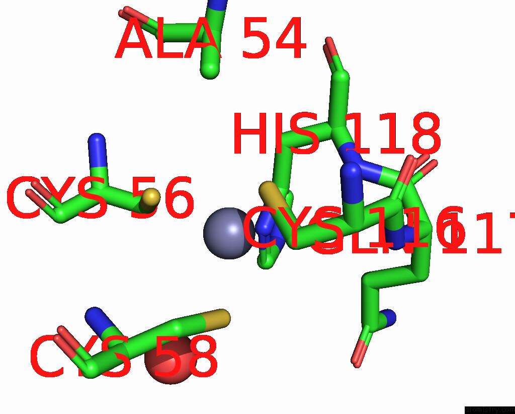
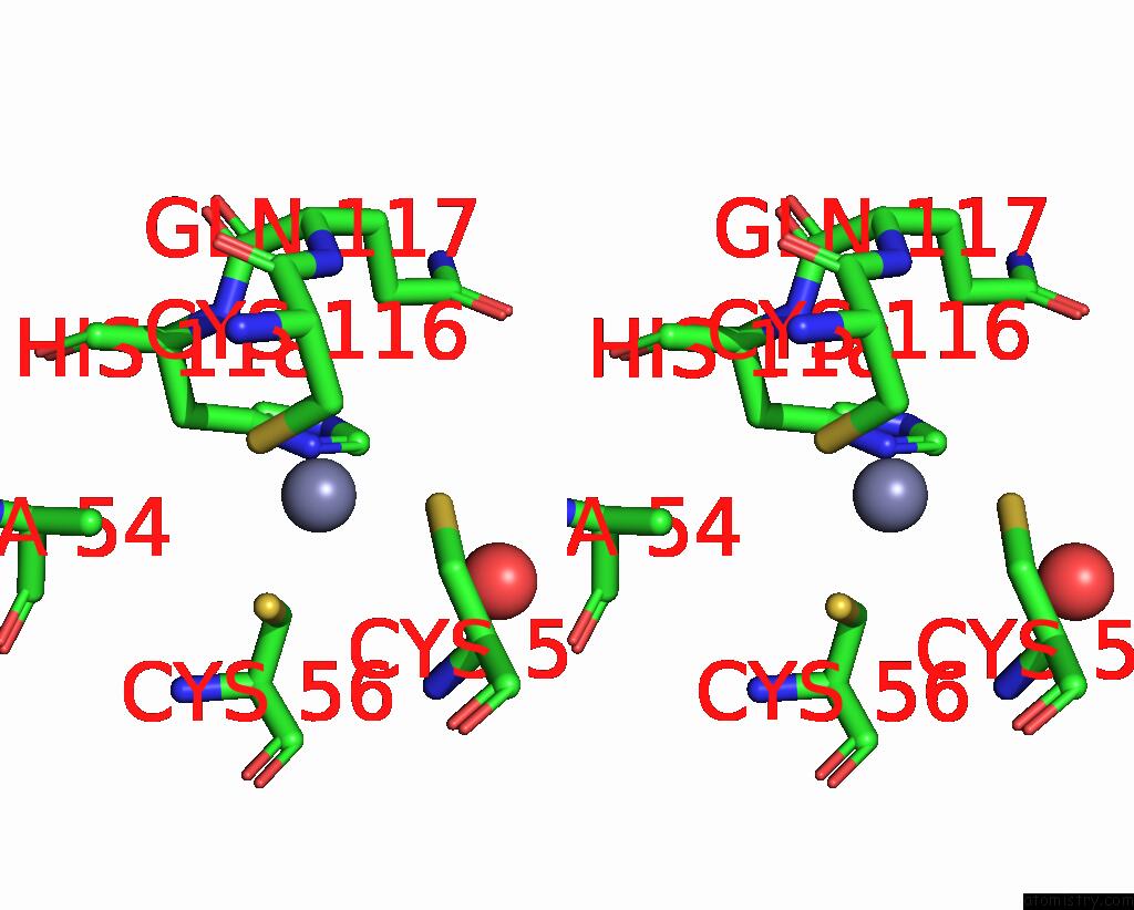
The binding sites of Zinc atom in the Group Deposition For Crystallographic Fragment Screening of Coxsackievirus A16 (G-10) 2A Protease -- Crystal Structure of Coxsackievirus A16 (G-10) 2A Protease in Complex with Z31602870 (A71EV2A-X0152)
(pdb code 7h2t). This binding sites where shown within
5.0 Angstroms radius around Zinc atom.
In total only one binding site of Zinc was determined in the Group Deposition For Crystallographic Fragment Screening of Coxsackievirus A16 (G-10) 2A Protease -- Crystal Structure of Coxsackievirus A16 (G-10) 2A Protease in Complex with Z31602870 (A71EV2A-X0152), PDB code: 7h2t:
In total only one binding site of Zinc was determined in the Group Deposition For Crystallographic Fragment Screening of Coxsackievirus A16 (G-10) 2A Protease -- Crystal Structure of Coxsackievirus A16 (G-10) 2A Protease in Complex with Z31602870 (A71EV2A-X0152), PDB code: 7h2t:
Zinc binding site 1 out of 1 in 7h2t
Go back to
Zinc binding site 1 out
of 1 in the Group Deposition For Crystallographic Fragment Screening of Coxsackievirus A16 (G-10) 2A Protease -- Crystal Structure of Coxsackievirus A16 (G-10) 2A Protease in Complex with Z31602870 (A71EV2A-X0152)
Mono view
Stereo pair view
Mono view
Stereo pair view
A full contact list of Zinc with other atoms in the Zn binding
site number 1 of Group Deposition For Crystallographic Fragment Screening of Coxsackievirus A16 (G-10) 2A Protease -- Crystal Structure of Coxsackievirus A16 (G-10) 2A Protease in Complex with Z31602870 (A71EV2A-X0152) within 5.0Å range:
|
Reference:
R.M.Lithgo,
M.Fairhead,
L.Koekemoer,
B.H.Balcomb,
E.Capkin,
A.V.Chandran,
M.Golding,
A.S.Godoy,
J.C.Aschenbrenner,
P.G.Marples,
X.Ni,
W.Thompson,
C.W.E.Tomlinson,
C.Wild,
M.Winokan,
M.-A.E.Xavier,
D.Fearon,
F.Von Delft.
Group Deposition For Crystallographic Fragment Screening of Coxsackievirus A16 (G-10) 2A Protease To Be Published.
Page generated: Tue Oct 29 20:43:50 2024
Last articles
Zn in 9MJ5Zn in 9HNW
Zn in 9G0L
Zn in 9FNE
Zn in 9DZN
Zn in 9E0I
Zn in 9D32
Zn in 9DAK
Zn in 8ZXC
Zn in 8ZUF