Zinc »
PDB 7cq4-7d3x »
7d2s »
Zinc in PDB 7d2s: Crystal Structure of RSU1/PINCH1_LIM5C Complex
Protein crystallography data
The structure of Crystal Structure of RSU1/PINCH1_LIM5C Complex, PDB code: 7d2s
was solved by
H.Yang,
Z.Wei,
Y.Cong,
with X-Ray Crystallography technique. A brief refinement statistics is given in the table below:
| Resolution Low / High (Å) | 50.00 / 1.65 |
| Space group | I 4 |
| Cell size a, b, c (Å), α, β, γ (°) | 124.596, 124.596, 50.518, 90, 90, 90 |
| R / Rfree (%) | 16.6 / 18.5 |
Zinc Binding Sites:
The binding sites of Zinc atom in the Crystal Structure of RSU1/PINCH1_LIM5C Complex
(pdb code 7d2s). This binding sites where shown within
5.0 Angstroms radius around Zinc atom.
In total 2 binding sites of Zinc where determined in the Crystal Structure of RSU1/PINCH1_LIM5C Complex, PDB code: 7d2s:
Jump to Zinc binding site number: 1; 2;
In total 2 binding sites of Zinc where determined in the Crystal Structure of RSU1/PINCH1_LIM5C Complex, PDB code: 7d2s:
Jump to Zinc binding site number: 1; 2;
Zinc binding site 1 out of 2 in 7d2s
Go back to
Zinc binding site 1 out
of 2 in the Crystal Structure of RSU1/PINCH1_LIM5C Complex
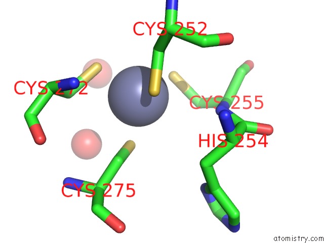
Mono view
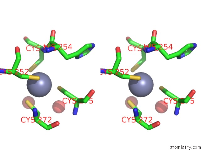
Stereo pair view
Mono view
Stereo pair view
A full contact list of Zinc with other atoms in the Zn binding
site number 1 of Crystal Structure of RSU1/PINCH1_LIM5C Complex within 5.0Å range:
|
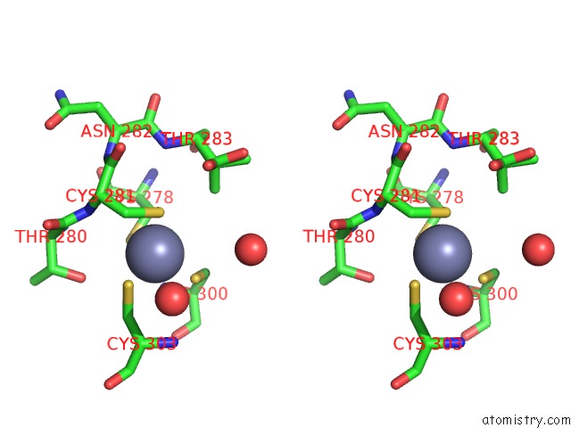
Zinc binding site 2 out of 2 in 7d2s
Go back to
Zinc binding site 2 out
of 2 in the Crystal Structure of RSU1/PINCH1_LIM5C Complex
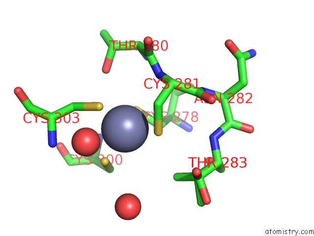
Mono view
Stereo pair view
Mono view
Stereo pair view
A full contact list of Zinc with other atoms in the Zn binding
site number 2 of Crystal Structure of RSU1/PINCH1_LIM5C Complex within 5.0Å range:
|
Reference:
H.Yang,
L.Lin,
K.Sun,
T.Zhang,
W.Chen,
L.Li,
Y.Xie,
C.Wu,
Z.Wei,
C.Yu.
Complex Structures of RSU1 and PINCH1 Reveal A Regulatory Mechanism of the Ilk/Pinch/Parvin Complex For F-Actin Dynamics. Elife V. 10 2021.
ISSN: ESSN 2050-084X
PubMed: 33587032
DOI: 10.7554/ELIFE.64395
Page generated: Tue Oct 29 18:36:22 2024
ISSN: ESSN 2050-084X
PubMed: 33587032
DOI: 10.7554/ELIFE.64395
Last articles
Zn in 9JYWZn in 9IR4
Zn in 9IR3
Zn in 9GMX
Zn in 9GMW
Zn in 9JEJ
Zn in 9ERF
Zn in 9ERE
Zn in 9EGV
Zn in 9EGW