Zinc »
PDB 7cgu-7cq3 »
7cph »
Zinc in PDB 7cph: Crystal Structure of Trna Adenosine Deaminase From Bacillus Subtilis
Enzymatic activity of Crystal Structure of Trna Adenosine Deaminase From Bacillus Subtilis
All present enzymatic activity of Crystal Structure of Trna Adenosine Deaminase From Bacillus Subtilis:
3.5.4.33;
3.5.4.33;
Protein crystallography data
The structure of Crystal Structure of Trna Adenosine Deaminase From Bacillus Subtilis, PDB code: 7cph
was solved by
V.Gaded,
J.Mariam,
R.Anand,
with X-Ray Crystallography technique. A brief refinement statistics is given in the table below:
| Resolution Low / High (Å) | 38.19 / 2.30 |
| Space group | P 21 21 21 |
| Cell size a, b, c (Å), α, β, γ (°) | 38.53, 41.25, 202.08, 90, 90, 90 |
| R / Rfree (%) | 17.7 / 24 |
Zinc Binding Sites:
The binding sites of Zinc atom in the Crystal Structure of Trna Adenosine Deaminase From Bacillus Subtilis
(pdb code 7cph). This binding sites where shown within
5.0 Angstroms radius around Zinc atom.
In total 2 binding sites of Zinc where determined in the Crystal Structure of Trna Adenosine Deaminase From Bacillus Subtilis, PDB code: 7cph:
Jump to Zinc binding site number: 1; 2;
In total 2 binding sites of Zinc where determined in the Crystal Structure of Trna Adenosine Deaminase From Bacillus Subtilis, PDB code: 7cph:
Jump to Zinc binding site number: 1; 2;
Zinc binding site 1 out of 2 in 7cph
Go back to
Zinc binding site 1 out
of 2 in the Crystal Structure of Trna Adenosine Deaminase From Bacillus Subtilis
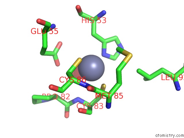
Mono view
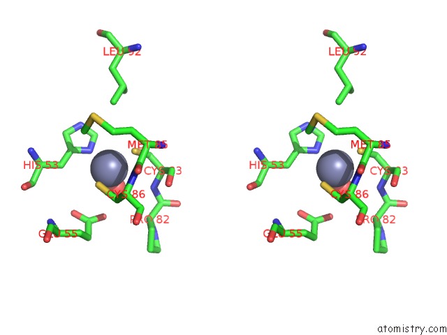
Stereo pair view
Mono view
Stereo pair view
A full contact list of Zinc with other atoms in the Zn binding
site number 1 of Crystal Structure of Trna Adenosine Deaminase From Bacillus Subtilis within 5.0Å range:
|
Zinc binding site 2 out of 2 in 7cph
Go back to
Zinc binding site 2 out
of 2 in the Crystal Structure of Trna Adenosine Deaminase From Bacillus Subtilis
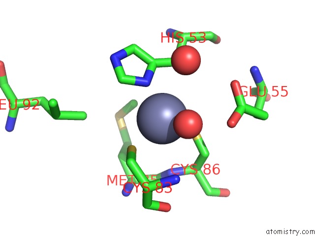
Mono view
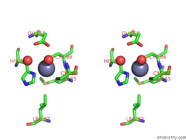
Stereo pair view
Mono view
Stereo pair view
A full contact list of Zinc with other atoms in the Zn binding
site number 2 of Crystal Structure of Trna Adenosine Deaminase From Bacillus Subtilis within 5.0Å range:
|
Reference:
J.Mariam,
A.Hoskere Ashoka,
V.Gaded,
F.Ali,
H.Malvi,
A.Das,
R.Anand.
Deciphering Protein Microenvironment By Using A Cysteine Specific Switch-on Fluorescent Probe. Org.Biomol.Chem. 2021.
ISSN: ESSN 1477-0539
PubMed: 34037063
DOI: 10.1039/D1OB00698C
Page generated: Tue Oct 29 18:23:27 2024
ISSN: ESSN 1477-0539
PubMed: 34037063
DOI: 10.1039/D1OB00698C
Last articles
Zn in 9JYWZn in 9IR4
Zn in 9IR3
Zn in 9GMX
Zn in 9GMW
Zn in 9JEJ
Zn in 9ERF
Zn in 9ERE
Zn in 9EGV
Zn in 9EGW