Zinc »
PDB 7auw-7b83 »
7b4b »
Zinc in PDB 7b4b: Structural Basis of Reactivation of Oncogenic P53 Mutants By A Small Molecule: Methylene Quinuclidinone (Mq). Human P53DBD-R273C Mutant Bound to Mq: R273C-Mq (I)
Protein crystallography data
The structure of Structural Basis of Reactivation of Oncogenic P53 Mutants By A Small Molecule: Methylene Quinuclidinone (Mq). Human P53DBD-R273C Mutant Bound to Mq: R273C-Mq (I), PDB code: 7b4b
was solved by
O.Degtjarik,
H.Rozenberg,
Z.Shakked,
with X-Ray Crystallography technique. A brief refinement statistics is given in the table below:
| Resolution Low / High (Å) | 36.46 / 1.76 |
| Space group | P 1 21 1 |
| Cell size a, b, c (Å), α, β, γ (°) | 68.73, 70.686, 85.107, 90, 90.29, 90 |
| R / Rfree (%) | 18 / 21.8 |
Zinc Binding Sites:
The binding sites of Zinc atom in the Structural Basis of Reactivation of Oncogenic P53 Mutants By A Small Molecule: Methylene Quinuclidinone (Mq). Human P53DBD-R273C Mutant Bound to Mq: R273C-Mq (I)
(pdb code 7b4b). This binding sites where shown within
5.0 Angstroms radius around Zinc atom.
In total 4 binding sites of Zinc where determined in the Structural Basis of Reactivation of Oncogenic P53 Mutants By A Small Molecule: Methylene Quinuclidinone (Mq). Human P53DBD-R273C Mutant Bound to Mq: R273C-Mq (I), PDB code: 7b4b:
Jump to Zinc binding site number: 1; 2; 3; 4;
In total 4 binding sites of Zinc where determined in the Structural Basis of Reactivation of Oncogenic P53 Mutants By A Small Molecule: Methylene Quinuclidinone (Mq). Human P53DBD-R273C Mutant Bound to Mq: R273C-Mq (I), PDB code: 7b4b:
Jump to Zinc binding site number: 1; 2; 3; 4;
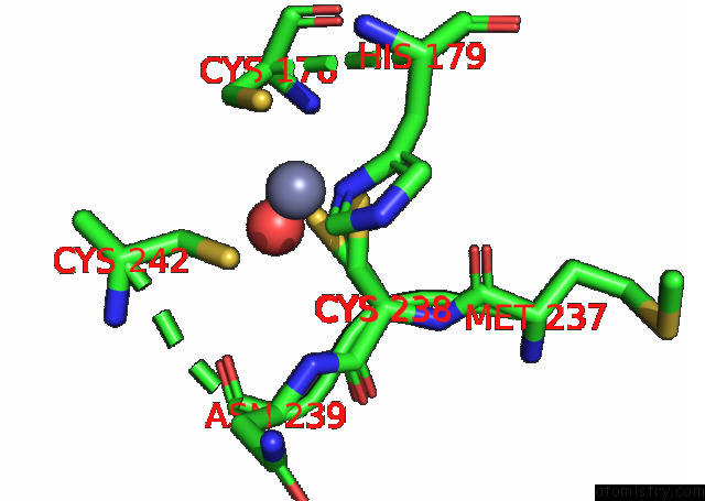
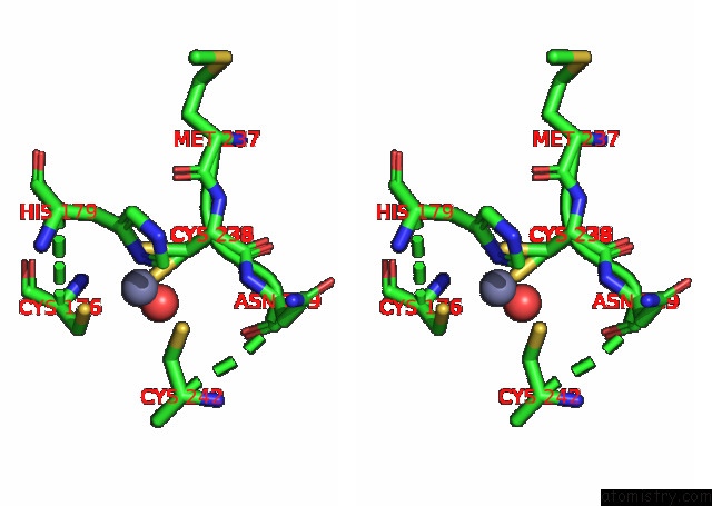
Zinc binding site 1 out of 4 in 7b4b
Go back to
Zinc binding site 1 out
of 4 in the Structural Basis of Reactivation of Oncogenic P53 Mutants By A Small Molecule: Methylene Quinuclidinone (Mq). Human P53DBD-R273C Mutant Bound to Mq: R273C-Mq (I)
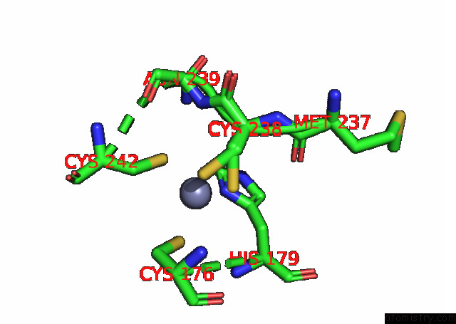
Mono view
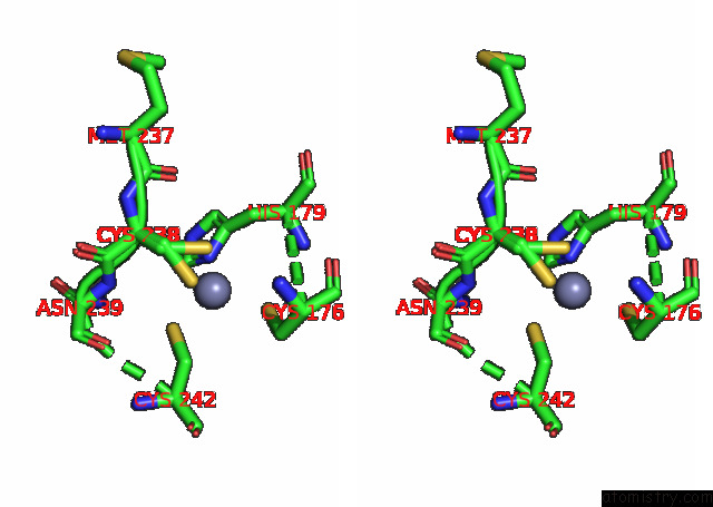
Stereo pair view
Mono view
Stereo pair view
A full contact list of Zinc with other atoms in the Zn binding
site number 1 of Structural Basis of Reactivation of Oncogenic P53 Mutants By A Small Molecule: Methylene Quinuclidinone (Mq). Human P53DBD-R273C Mutant Bound to Mq: R273C-Mq (I) within 5.0Å range:
|
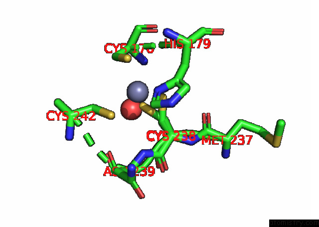
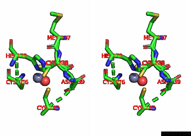
Zinc binding site 2 out of 4 in 7b4b
Go back to
Zinc binding site 2 out
of 4 in the Structural Basis of Reactivation of Oncogenic P53 Mutants By A Small Molecule: Methylene Quinuclidinone (Mq). Human P53DBD-R273C Mutant Bound to Mq: R273C-Mq (I)
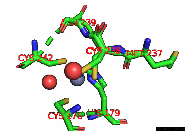
Mono view
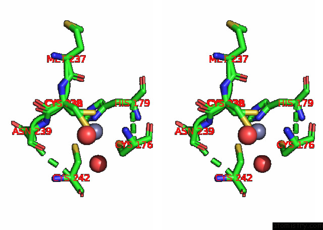
Stereo pair view
Mono view
Stereo pair view
A full contact list of Zinc with other atoms in the Zn binding
site number 2 of Structural Basis of Reactivation of Oncogenic P53 Mutants By A Small Molecule: Methylene Quinuclidinone (Mq). Human P53DBD-R273C Mutant Bound to Mq: R273C-Mq (I) within 5.0Å range:
|
Zinc binding site 3 out of 4 in 7b4b
Go back to
Zinc binding site 3 out
of 4 in the Structural Basis of Reactivation of Oncogenic P53 Mutants By A Small Molecule: Methylene Quinuclidinone (Mq). Human P53DBD-R273C Mutant Bound to Mq: R273C-Mq (I)
Mono view
Stereo pair view
Mono view
Stereo pair view
A full contact list of Zinc with other atoms in the Zn binding
site number 3 of Structural Basis of Reactivation of Oncogenic P53 Mutants By A Small Molecule: Methylene Quinuclidinone (Mq). Human P53DBD-R273C Mutant Bound to Mq: R273C-Mq (I) within 5.0Å range:
|
Zinc binding site 4 out of 4 in 7b4b
Go back to
Zinc binding site 4 out
of 4 in the Structural Basis of Reactivation of Oncogenic P53 Mutants By A Small Molecule: Methylene Quinuclidinone (Mq). Human P53DBD-R273C Mutant Bound to Mq: R273C-Mq (I)
Mono view
Stereo pair view
Mono view
Stereo pair view
A full contact list of Zinc with other atoms in the Zn binding
site number 4 of Structural Basis of Reactivation of Oncogenic P53 Mutants By A Small Molecule: Methylene Quinuclidinone (Mq). Human P53DBD-R273C Mutant Bound to Mq: R273C-Mq (I) within 5.0Å range:
|
Reference:
O.Degtjarik,
D.Golovenko,
Y.Diskin-Posner,
L.Abrahmsen,
H.Rozenberg,
Z.Shakked.
P53 Structure 5 To Be Published.
Page generated: Tue Oct 29 17:25:14 2024
Last articles
Zn in 9JYWZn in 9IR4
Zn in 9IR3
Zn in 9GMX
Zn in 9GMW
Zn in 9JEJ
Zn in 9ERF
Zn in 9ERE
Zn in 9EGV
Zn in 9EGW