Zinc »
PDB 6xty-6y77 »
6xwg »
Zinc in PDB 6xwg: Crystal Structure of the Human Rxr/Rar Dna-Binding Domain Heterodimer Bound to the Human RARB2 DR5 Response Element
Protein crystallography data
The structure of Crystal Structure of the Human Rxr/Rar Dna-Binding Domain Heterodimer Bound to the Human RARB2 DR5 Response Element, PDB code: 6xwg
was solved by
A.G.Mcewen,
P.Poussin-Courmontagne,
C.Peluso-Iltis,
N.Rochel,
with X-Ray Crystallography technique. A brief refinement statistics is given in the table below:
| Resolution Low / High (Å) | 26.51 / 2.40 |
| Space group | P 43 21 2 |
| Cell size a, b, c (Å), α, β, γ (°) | 56.714, 56.714, 238.364, 90.00, 90.00, 90.00 |
| R / Rfree (%) | 19.1 / 20.7 |
Other elements in 6xwg:
The structure of Crystal Structure of the Human Rxr/Rar Dna-Binding Domain Heterodimer Bound to the Human RARB2 DR5 Response Element also contains other interesting chemical elements:
| Chlorine | (Cl) | 1 atom |
Zinc Binding Sites:
The binding sites of Zinc atom in the Crystal Structure of the Human Rxr/Rar Dna-Binding Domain Heterodimer Bound to the Human RARB2 DR5 Response Element
(pdb code 6xwg). This binding sites where shown within
5.0 Angstroms radius around Zinc atom.
In total 4 binding sites of Zinc where determined in the Crystal Structure of the Human Rxr/Rar Dna-Binding Domain Heterodimer Bound to the Human RARB2 DR5 Response Element, PDB code: 6xwg:
Jump to Zinc binding site number: 1; 2; 3; 4;
In total 4 binding sites of Zinc where determined in the Crystal Structure of the Human Rxr/Rar Dna-Binding Domain Heterodimer Bound to the Human RARB2 DR5 Response Element, PDB code: 6xwg:
Jump to Zinc binding site number: 1; 2; 3; 4;
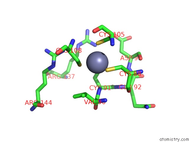
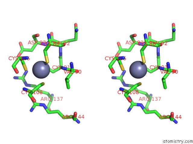
Zinc binding site 1 out of 4 in 6xwg
Go back to
Zinc binding site 1 out
of 4 in the Crystal Structure of the Human Rxr/Rar Dna-Binding Domain Heterodimer Bound to the Human RARB2 DR5 Response Element
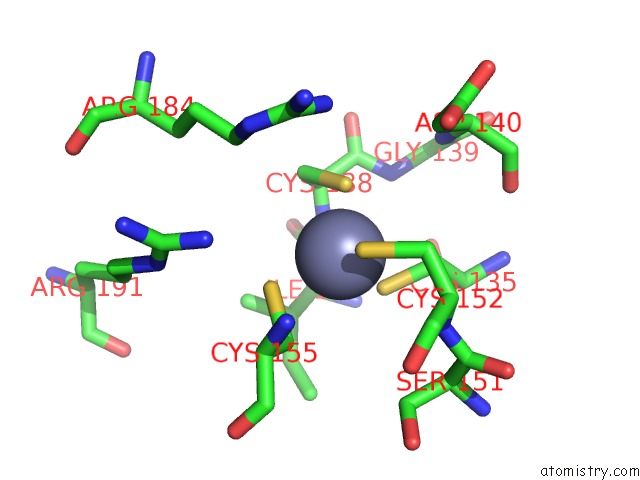
Mono view
Stereo pair view
Mono view
Stereo pair view
A full contact list of Zinc with other atoms in the Zn binding
site number 1 of Crystal Structure of the Human Rxr/Rar Dna-Binding Domain Heterodimer Bound to the Human RARB2 DR5 Response Element within 5.0Å range:
|
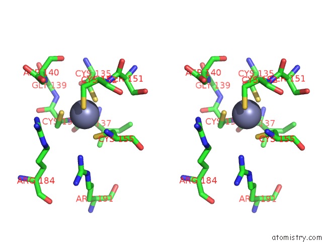
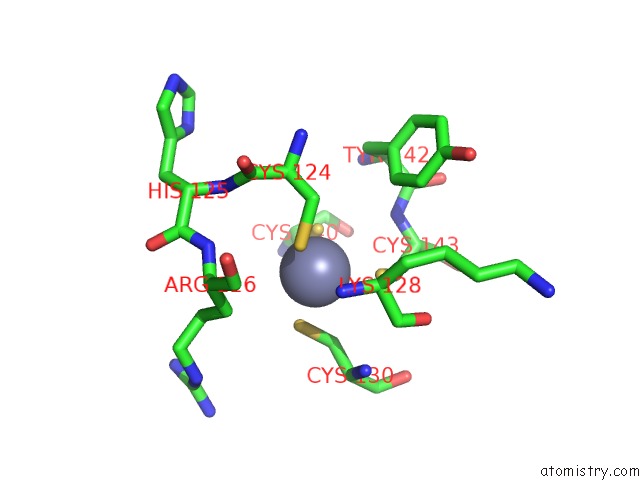
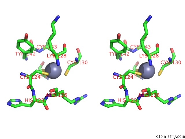
Zinc binding site 2 out of 4 in 6xwg
Go back to
Zinc binding site 2 out
of 4 in the Crystal Structure of the Human Rxr/Rar Dna-Binding Domain Heterodimer Bound to the Human RARB2 DR5 Response Element
Mono view
Stereo pair view
Mono view
Stereo pair view
A full contact list of Zinc with other atoms in the Zn binding
site number 2 of Crystal Structure of the Human Rxr/Rar Dna-Binding Domain Heterodimer Bound to the Human RARB2 DR5 Response Element within 5.0Å range:
|
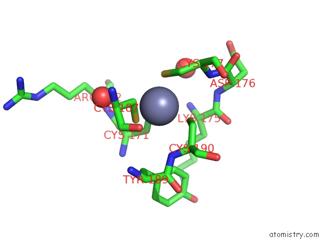
Zinc binding site 3 out of 4 in 6xwg
Go back to
Zinc binding site 3 out
of 4 in the Crystal Structure of the Human Rxr/Rar Dna-Binding Domain Heterodimer Bound to the Human RARB2 DR5 Response Element
Mono view
Stereo pair view
Mono view
Stereo pair view
A full contact list of Zinc with other atoms in the Zn binding
site number 3 of Crystal Structure of the Human Rxr/Rar Dna-Binding Domain Heterodimer Bound to the Human RARB2 DR5 Response Element within 5.0Å range:
|
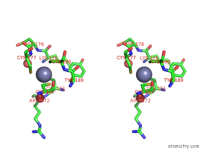
Zinc binding site 4 out of 4 in 6xwg
Go back to
Zinc binding site 4 out
of 4 in the Crystal Structure of the Human Rxr/Rar Dna-Binding Domain Heterodimer Bound to the Human RARB2 DR5 Response Element
Mono view
Stereo pair view
Mono view
Stereo pair view
A full contact list of Zinc with other atoms in the Zn binding
site number 4 of Crystal Structure of the Human Rxr/Rar Dna-Binding Domain Heterodimer Bound to the Human RARB2 DR5 Response Element within 5.0Å range:
|
Reference:
J.Osz,
A.G.Mcewen,
M.Bourguet,
F.Przybilla,
C.Peluso-Iltis,
P.Poussin-Courmontagne,
Y.Mely,
S.Cianferani,
C.M.Jeffries,
D.I.Svergun,
N.Rochel.
Structural Basis For Dna Recognition and Allosteric Control of the Retinoic Acid Receptors Rar-Rxr. Nucleic Acids Res. V. 48 9969 2020.
ISSN: ESSN 1362-4962
PubMed: 32974652
DOI: 10.1093/NAR/GKAA697
Page generated: Tue Oct 29 11:16:01 2024
ISSN: ESSN 1362-4962
PubMed: 32974652
DOI: 10.1093/NAR/GKAA697
Last articles
Zn in 9JYWZn in 9IR4
Zn in 9IR3
Zn in 9GMX
Zn in 9GMW
Zn in 9JEJ
Zn in 9ERF
Zn in 9ERE
Zn in 9EGV
Zn in 9EGW