Zinc »
PDB 6rok-6rwg »
6rok »
Zinc in PDB 6rok: The Crystal Structure of A Complex Between the Llfpg Protein, A Thf- Dna and An Inhibitor
Enzymatic activity of The Crystal Structure of A Complex Between the Llfpg Protein, A Thf- Dna and An Inhibitor
All present enzymatic activity of The Crystal Structure of A Complex Between the Llfpg Protein, A Thf- Dna and An Inhibitor:
3.2.2.23; 4.2.99.18;
3.2.2.23; 4.2.99.18;
Protein crystallography data
The structure of The Crystal Structure of A Complex Between the Llfpg Protein, A Thf- Dna and An Inhibitor, PDB code: 6rok
was solved by
F.Coste,
S.Goffinont,
B.Castaing,
with X-Ray Crystallography technique. A brief refinement statistics is given in the table below:
| Resolution Low / High (Å) | 47.84 / 1.95 |
| Space group | P 41 21 2 |
| Cell size a, b, c (Å), α, β, γ (°) | 91.633, 91.633, 141.858, 90.00, 90.00, 90.00 |
| R / Rfree (%) | 16.8 / 19.6 |
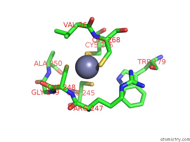
Zinc Binding Sites:
The binding sites of Zinc atom in the The Crystal Structure of A Complex Between the Llfpg Protein, A Thf- Dna and An Inhibitor
(pdb code 6rok). This binding sites where shown within
5.0 Angstroms radius around Zinc atom.
In total only one binding site of Zinc was determined in the The Crystal Structure of A Complex Between the Llfpg Protein, A Thf- Dna and An Inhibitor, PDB code: 6rok:
In total only one binding site of Zinc was determined in the The Crystal Structure of A Complex Between the Llfpg Protein, A Thf- Dna and An Inhibitor, PDB code: 6rok:
Zinc binding site 1 out of 1 in 6rok
Go back to
Zinc binding site 1 out
of 1 in the The Crystal Structure of A Complex Between the Llfpg Protein, A Thf- Dna and An Inhibitor
Mono view
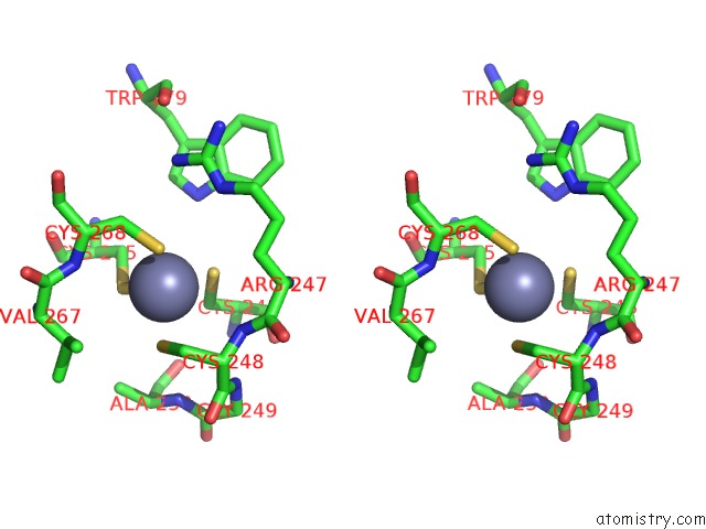
Stereo pair view
Mono view
Stereo pair view
A full contact list of Zinc with other atoms in the Zn binding
site number 1 of The Crystal Structure of A Complex Between the Llfpg Protein, A Thf- Dna and An Inhibitor within 5.0Å range:
|
Reference:
C.Rieux,
S.Goffinont,
F.Coste,
Z.Tber,
J.Cros,
V.Roy,
M.Guerin,
V.Gaudon,
S.Bourg,
A.Biela,
V.Aucagne,
L.Agrofoglio,
N.Garnier,
B.Castaing.
Thiopurine Derivative-Induced Fpg/Nei Dna Glycosylase Inhibition: Structural, Dynamic and Functional Insights. Int J Mol Sci V. 21 2020.
ISSN: ESSN 1422-0067
PubMed: 32192183
DOI: 10.3390/IJMS21062058
Page generated: Tue Oct 29 06:41:51 2024
ISSN: ESSN 1422-0067
PubMed: 32192183
DOI: 10.3390/IJMS21062058
Last articles
Zn in 9JYWZn in 9IR4
Zn in 9IR3
Zn in 9GMX
Zn in 9GMW
Zn in 9JEJ
Zn in 9ERF
Zn in 9ERE
Zn in 9EGV
Zn in 9EGW