Zinc »
PDB 6qci-6qxu »
6qu1 »
Zinc in PDB 6qu1: Crystal Structure of the KAP1 Rbcc Domain in Complex with the SMARCAD1 CUE1 Domain at 3.7 Angstrom Resolution.
Enzymatic activity of Crystal Structure of the KAP1 Rbcc Domain in Complex with the SMARCAD1 CUE1 Domain at 3.7 Angstrom Resolution.
All present enzymatic activity of Crystal Structure of the KAP1 Rbcc Domain in Complex with the SMARCAD1 CUE1 Domain at 3.7 Angstrom Resolution.:
2.3.2.27; 3.6.4.12;
2.3.2.27; 3.6.4.12;
Protein crystallography data
The structure of Crystal Structure of the KAP1 Rbcc Domain in Complex with the SMARCAD1 CUE1 Domain at 3.7 Angstrom Resolution., PDB code: 6qu1
was solved by
J.A.Newman,
H.Aitkenhead,
A.Gavard,
M.Lim,
H.L.Williams,
J.Q.Svejstrup,
F.Von Delft,
C.H.Arrowsmith,
A.Edwards,
C.Bountra,
O.Gileadi,
with X-Ray Crystallography technique. A brief refinement statistics is given in the table below:
| Resolution Low / High (Å) | 29.43 / 3.70 |
| Space group | P 41 21 2 |
| Cell size a, b, c (Å), α, β, γ (°) | 64.496, 64.496, 287.986, 90.00, 90.00, 90.00 |
| R / Rfree (%) | 30 / 34.6 |
Zinc Binding Sites:
The binding sites of Zinc atom in the Crystal Structure of the KAP1 Rbcc Domain in Complex with the SMARCAD1 CUE1 Domain at 3.7 Angstrom Resolution.
(pdb code 6qu1). This binding sites where shown within
5.0 Angstroms radius around Zinc atom.
In total 4 binding sites of Zinc where determined in the Crystal Structure of the KAP1 Rbcc Domain in Complex with the SMARCAD1 CUE1 Domain at 3.7 Angstrom Resolution., PDB code: 6qu1:
Jump to Zinc binding site number: 1; 2; 3; 4;
In total 4 binding sites of Zinc where determined in the Crystal Structure of the KAP1 Rbcc Domain in Complex with the SMARCAD1 CUE1 Domain at 3.7 Angstrom Resolution., PDB code: 6qu1:
Jump to Zinc binding site number: 1; 2; 3; 4;
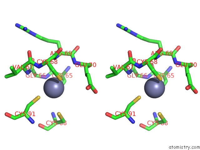
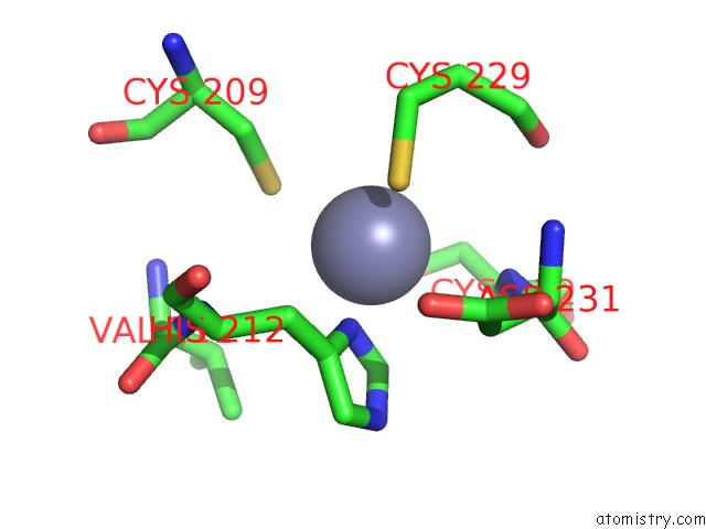
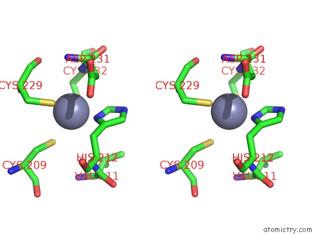
Zinc binding site 1 out of 4 in 6qu1
Go back to
Zinc binding site 1 out
of 4 in the Crystal Structure of the KAP1 Rbcc Domain in Complex with the SMARCAD1 CUE1 Domain at 3.7 Angstrom Resolution.
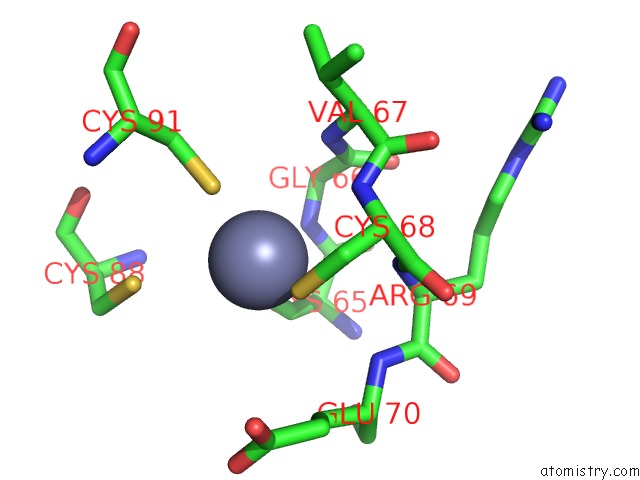
Mono view
Stereo pair view
Mono view
Stereo pair view
A full contact list of Zinc with other atoms in the Zn binding
site number 1 of Crystal Structure of the KAP1 Rbcc Domain in Complex with the SMARCAD1 CUE1 Domain at 3.7 Angstrom Resolution. within 5.0Å range:
|
Zinc binding site 2 out of 4 in 6qu1
Go back to
Zinc binding site 2 out
of 4 in the Crystal Structure of the KAP1 Rbcc Domain in Complex with the SMARCAD1 CUE1 Domain at 3.7 Angstrom Resolution.
Mono view
Stereo pair view
Mono view
Stereo pair view
A full contact list of Zinc with other atoms in the Zn binding
site number 2 of Crystal Structure of the KAP1 Rbcc Domain in Complex with the SMARCAD1 CUE1 Domain at 3.7 Angstrom Resolution. within 5.0Å range:
|
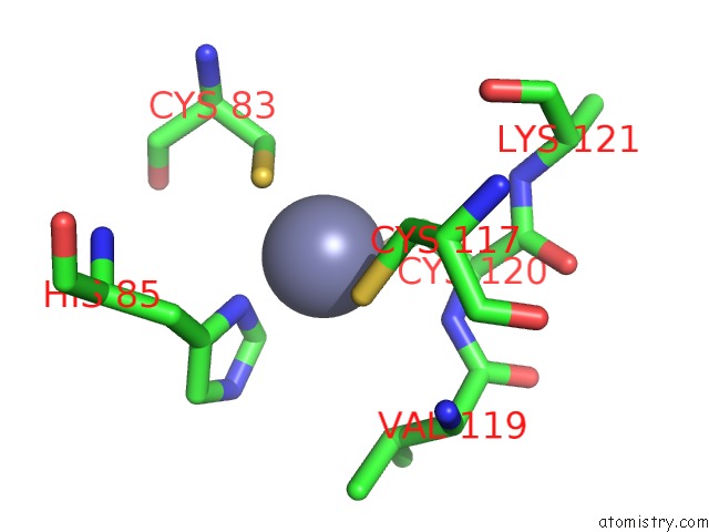
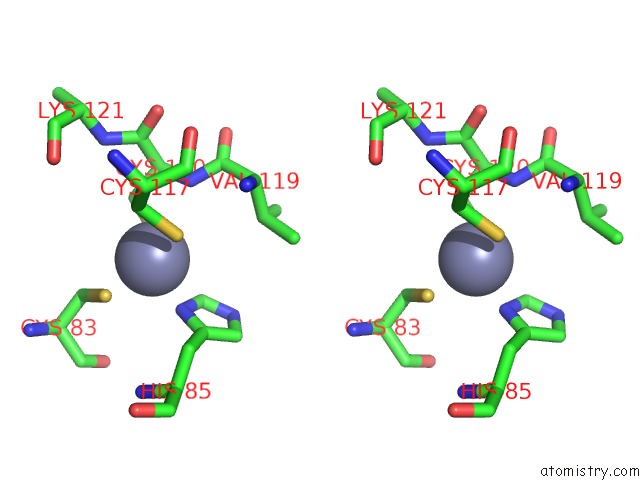
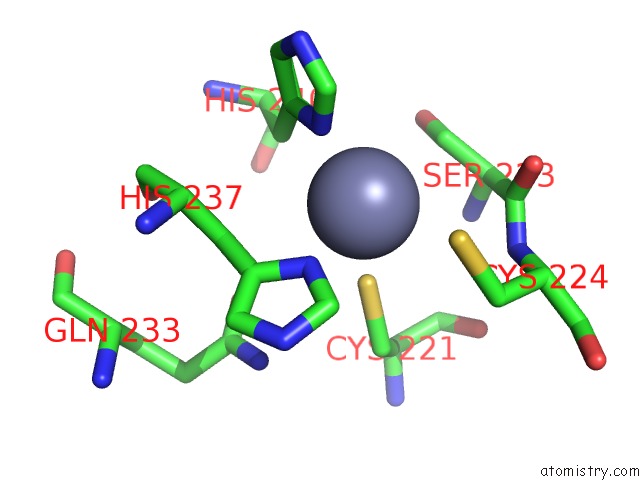
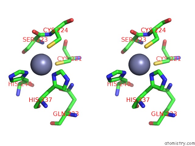
Zinc binding site 3 out of 4 in 6qu1
Go back to
Zinc binding site 3 out
of 4 in the Crystal Structure of the KAP1 Rbcc Domain in Complex with the SMARCAD1 CUE1 Domain at 3.7 Angstrom Resolution.
Mono view
Stereo pair view
Mono view
Stereo pair view
A full contact list of Zinc with other atoms in the Zn binding
site number 3 of Crystal Structure of the KAP1 Rbcc Domain in Complex with the SMARCAD1 CUE1 Domain at 3.7 Angstrom Resolution. within 5.0Å range:
|
Zinc binding site 4 out of 4 in 6qu1
Go back to
Zinc binding site 4 out
of 4 in the Crystal Structure of the KAP1 Rbcc Domain in Complex with the SMARCAD1 CUE1 Domain at 3.7 Angstrom Resolution.
Mono view
Stereo pair view
Mono view
Stereo pair view
A full contact list of Zinc with other atoms in the Zn binding
site number 4 of Crystal Structure of the KAP1 Rbcc Domain in Complex with the SMARCAD1 CUE1 Domain at 3.7 Angstrom Resolution. within 5.0Å range:
|
Reference:
M.Lim,
J.A.Newman,
H.L.Williams,
L.Masino,
H.Aitkenhead,
A.E.Gravard,
O.Gileadi,
J.Q.Svejstrup.
A Ubiquitin-Binding Domain That Binds A Structural Fold Distinct From That of Ubiquitin. Structure 2019.
ISSN: ISSN 0969-2126
PubMed: 31204252
DOI: 10.1016/J.STR.2019.05.003
Page generated: Tue Oct 29 05:46:15 2024
ISSN: ISSN 0969-2126
PubMed: 31204252
DOI: 10.1016/J.STR.2019.05.003
Last articles
Zn in 9JYWZn in 9IR4
Zn in 9IR3
Zn in 9GMX
Zn in 9GMW
Zn in 9JEJ
Zn in 9ERF
Zn in 9ERE
Zn in 9EGV
Zn in 9EGW