Zinc »
PDB 6ouc-6p3y »
6p1g »
Zinc in PDB 6p1g: Copper-Bound Pcuac Domain From PMOF2
Protein crystallography data
The structure of Copper-Bound Pcuac Domain From PMOF2, PDB code: 6p1g
was solved by
O.S.Fisher,
A.C.Rosenzweig,
with X-Ray Crystallography technique. A brief refinement statistics is given in the table below:
| Resolution Low / High (Å) | 35.85 / 2.05 |
| Space group | P 32 2 1 |
| Cell size a, b, c (Å), α, β, γ (°) | 89.350, 89.350, 60.070, 90.00, 90.00, 120.00 |
| R / Rfree (%) | 18.6 / 22.9 |
Other elements in 6p1g:
The structure of Copper-Bound Pcuac Domain From PMOF2 also contains other interesting chemical elements:
| Copper | (Cu) | 2 atoms |
Zinc Binding Sites:
The binding sites of Zinc atom in the Copper-Bound Pcuac Domain From PMOF2
(pdb code 6p1g). This binding sites where shown within
5.0 Angstroms radius around Zinc atom.
In total 2 binding sites of Zinc where determined in the Copper-Bound Pcuac Domain From PMOF2, PDB code: 6p1g:
Jump to Zinc binding site number: 1; 2;
In total 2 binding sites of Zinc where determined in the Copper-Bound Pcuac Domain From PMOF2, PDB code: 6p1g:
Jump to Zinc binding site number: 1; 2;
Zinc binding site 1 out of 2 in 6p1g
Go back to
Zinc binding site 1 out
of 2 in the Copper-Bound Pcuac Domain From PMOF2
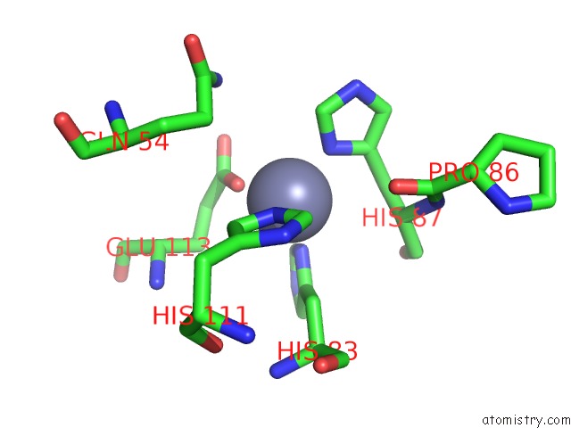
Mono view
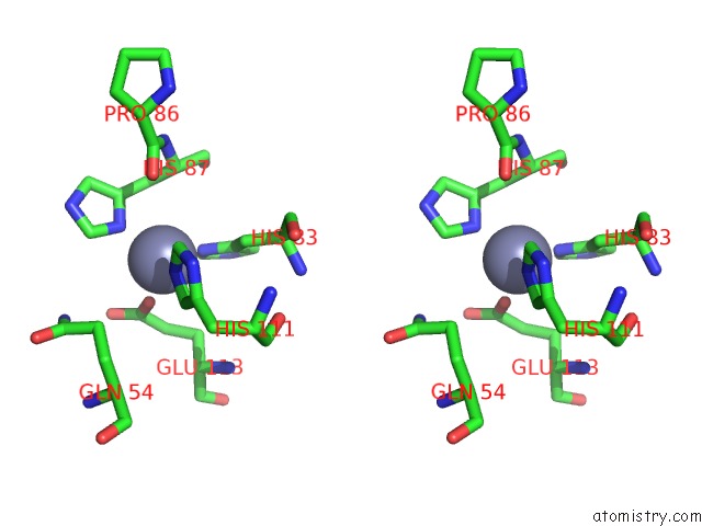
Stereo pair view
Mono view
Stereo pair view
A full contact list of Zinc with other atoms in the Zn binding
site number 1 of Copper-Bound Pcuac Domain From PMOF2 within 5.0Å range:
|
Zinc binding site 2 out of 2 in 6p1g
Go back to
Zinc binding site 2 out
of 2 in the Copper-Bound Pcuac Domain From PMOF2
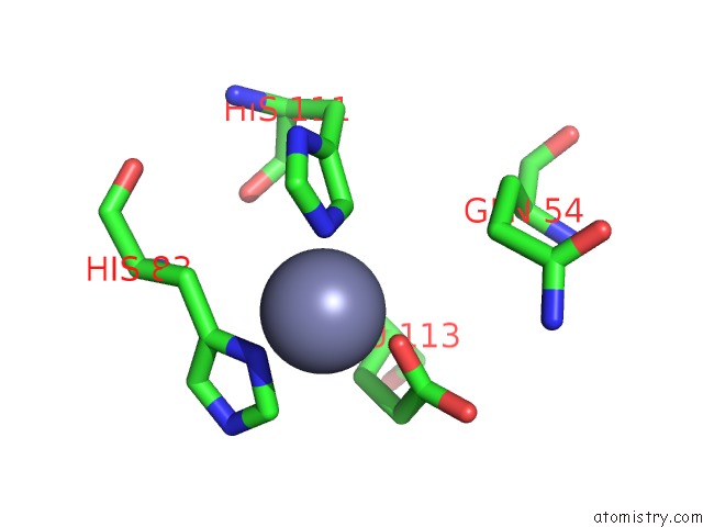
Mono view
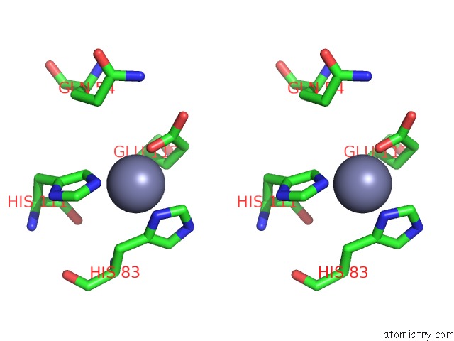
Stereo pair view
Mono view
Stereo pair view
A full contact list of Zinc with other atoms in the Zn binding
site number 2 of Copper-Bound Pcuac Domain From PMOF2 within 5.0Å range:
|
Reference:
O.S.Fisher,
M.R.Sendzik,
M.O.Ross,
T.J.Lawton,
B.M.Hoffman,
A.C.Rosenzweig.
Pcuac Domains From Methane-Oxidizing Bacteria Use A Histidine Brace to Bind Copper. J.Biol.Chem. V. 294 16351 2019.
ISSN: ESSN 1083-351X
PubMed: 31527086
DOI: 10.1074/JBC.RA119.010093
Page generated: Tue Oct 29 04:45:34 2024
ISSN: ESSN 1083-351X
PubMed: 31527086
DOI: 10.1074/JBC.RA119.010093
Last articles
Zn in 9JYWZn in 9IR4
Zn in 9IR3
Zn in 9GMX
Zn in 9GMW
Zn in 9JEJ
Zn in 9ERF
Zn in 9ERE
Zn in 9EGV
Zn in 9EGW