Zinc »
PDB 6a3n-6aem »
6a8z »
Zinc in PDB 6a8z: Crystal Structure of M1 Zinc Metallopeptidase From Deinococcus Radiodurans
Protein crystallography data
The structure of Crystal Structure of M1 Zinc Metallopeptidase From Deinococcus Radiodurans, PDB code: 6a8z
was solved by
R.Agrawal,
A.Kumar,
R.D.Makde,
with X-Ray Crystallography technique. A brief refinement statistics is given in the table below:
| Resolution Low / High (Å) | 30.25 / 2.05 |
| Space group | P 1 |
| Cell size a, b, c (Å), α, β, γ (°) | 52.371, 57.583, 69.579, 89.74, 82.30, 67.53 |
| R / Rfree (%) | 17.5 / 22 |
Other elements in 6a8z:
The structure of Crystal Structure of M1 Zinc Metallopeptidase From Deinococcus Radiodurans also contains other interesting chemical elements:
| Sodium | (Na) | 2 atoms |
Zinc Binding Sites:
The binding sites of Zinc atom in the Crystal Structure of M1 Zinc Metallopeptidase From Deinococcus Radiodurans
(pdb code 6a8z). This binding sites where shown within
5.0 Angstroms radius around Zinc atom.
In total 2 binding sites of Zinc where determined in the Crystal Structure of M1 Zinc Metallopeptidase From Deinococcus Radiodurans, PDB code: 6a8z:
Jump to Zinc binding site number: 1; 2;
In total 2 binding sites of Zinc where determined in the Crystal Structure of M1 Zinc Metallopeptidase From Deinococcus Radiodurans, PDB code: 6a8z:
Jump to Zinc binding site number: 1; 2;
Zinc binding site 1 out of 2 in 6a8z
Go back to
Zinc binding site 1 out
of 2 in the Crystal Structure of M1 Zinc Metallopeptidase From Deinococcus Radiodurans
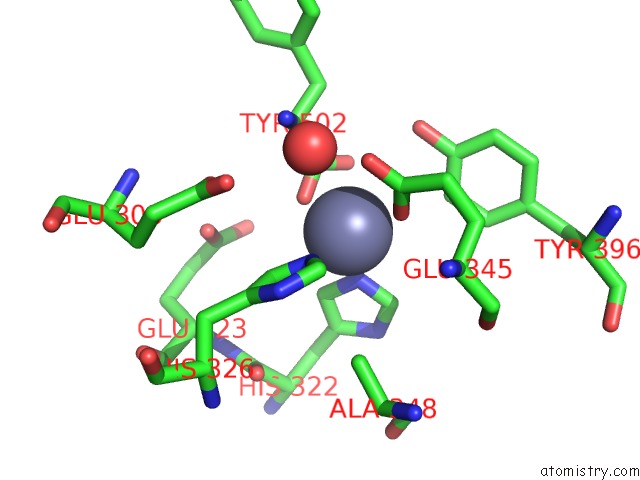
Mono view
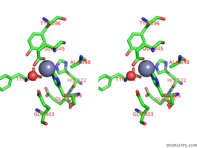
Stereo pair view
Mono view
Stereo pair view
A full contact list of Zinc with other atoms in the Zn binding
site number 1 of Crystal Structure of M1 Zinc Metallopeptidase From Deinococcus Radiodurans within 5.0Å range:
|
Zinc binding site 2 out of 2 in 6a8z
Go back to
Zinc binding site 2 out
of 2 in the Crystal Structure of M1 Zinc Metallopeptidase From Deinococcus Radiodurans
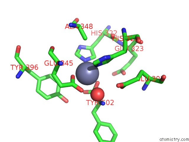
Mono view
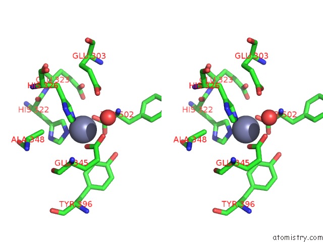
Stereo pair view
Mono view
Stereo pair view
A full contact list of Zinc with other atoms in the Zn binding
site number 2 of Crystal Structure of M1 Zinc Metallopeptidase From Deinococcus Radiodurans within 5.0Å range:
|
Reference:
R.Agrawal,
V.D.Goyal,
A.Kumar,
N.K.Gaur,
S.N.Jamdar,
A.Kumar,
R.D.Makde.
Two-Domain Aminopeptidase of M1 Family: Structural Features For Substrate Binding and Gating in Absence of C-Terminal Domain. J.Struct.Biol. V. 208 51 2019.
ISSN: ESSN 1095-8657
PubMed: 31351924
DOI: 10.1016/J.JSB.2019.07.010
Page generated: Mon Oct 28 17:26:35 2024
ISSN: ESSN 1095-8657
PubMed: 31351924
DOI: 10.1016/J.JSB.2019.07.010
Last articles
Zn in 9JYWZn in 9IR4
Zn in 9IR3
Zn in 9GMX
Zn in 9GMW
Zn in 9JEJ
Zn in 9ERF
Zn in 9ERE
Zn in 9EGV
Zn in 9EGW