Zinc »
PDB 6a57-6agg »
6a83 »
Zinc in PDB 6a83: Crystal Structure of the C-Terminal Periplasmic Domain of Eceptc From Escherichia Coli Complex with Zn
Protein crystallography data
The structure of Crystal Structure of the C-Terminal Periplasmic Domain of Eceptc From Escherichia Coli Complex with Zn, PDB code: 6a83
was solved by
Y.Q.Zhao,
Y.J.Gu,
W.Cheng,
with X-Ray Crystallography technique. A brief refinement statistics is given in the table below:
| Resolution Low / High (Å) | 47.69 / 2.60 |
| Space group | P 32 2 1 |
| Cell size a, b, c (Å), α, β, γ (°) | 103.568, 103.568, 122.294, 90.00, 90.00, 120.00 |
| R / Rfree (%) | 21.2 / 23.7 |
Other elements in 6a83:
The structure of Crystal Structure of the C-Terminal Periplasmic Domain of Eceptc From Escherichia Coli Complex with Zn also contains other interesting chemical elements:
| Sodium | (Na) | 1 atom |
Zinc Binding Sites:
The binding sites of Zinc atom in the Crystal Structure of the C-Terminal Periplasmic Domain of Eceptc From Escherichia Coli Complex with Zn
(pdb code 6a83). This binding sites where shown within
5.0 Angstroms radius around Zinc atom.
In total only one binding site of Zinc was determined in the Crystal Structure of the C-Terminal Periplasmic Domain of Eceptc From Escherichia Coli Complex with Zn, PDB code: 6a83:
In total only one binding site of Zinc was determined in the Crystal Structure of the C-Terminal Periplasmic Domain of Eceptc From Escherichia Coli Complex with Zn, PDB code: 6a83:
Zinc binding site 1 out of 1 in 6a83
Go back to
Zinc binding site 1 out
of 1 in the Crystal Structure of the C-Terminal Periplasmic Domain of Eceptc From Escherichia Coli Complex with Zn
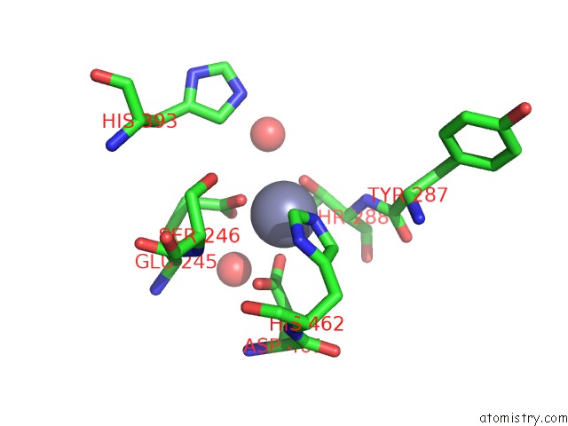
Mono view
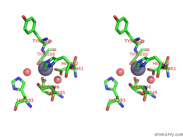
Stereo pair view
Mono view
Stereo pair view
A full contact list of Zinc with other atoms in the Zn binding
site number 1 of Crystal Structure of the C-Terminal Periplasmic Domain of Eceptc From Escherichia Coli Complex with Zn within 5.0Å range:
|
Reference:
Y.Zhao,
Q.Meng,
Y.Lai,
L.Wang,
D.Zhou,
C.Dou,
Y.Gu,
C.Nie,
Y.Wei,
W.Cheng.
Structural and Mechanistic Insights Into Polymyxin Resistance Mediated By Eptc Originating From Escherichia Coli. Febs J. V. 286 750 2019.
ISSN: ISSN 1742-4658
PubMed: 30537137
DOI: 10.1111/FEBS.14719
Page generated: Mon Oct 28 17:24:44 2024
ISSN: ISSN 1742-4658
PubMed: 30537137
DOI: 10.1111/FEBS.14719
Last articles
Zn in 9MJ5Zn in 9HNW
Zn in 9G0L
Zn in 9FNE
Zn in 9DZN
Zn in 9E0I
Zn in 9D32
Zn in 9DAK
Zn in 8ZXC
Zn in 8ZUF