Zinc »
PDB 5xna-5y1q »
5xxe »
Zinc in PDB 5xxe: Crystal Structure of POZ1 and TPZ1
Protein crystallography data
The structure of Crystal Structure of POZ1 and TPZ1, PDB code: 5xxe
was solved by
J.Xue,
H.Chen,
J.Wu,
M.Lei,
with X-Ray Crystallography technique. A brief refinement statistics is given in the table below:
| Resolution Low / High (Å) | 50.00 / 2.50 |
| Space group | P 21 21 21 |
| Cell size a, b, c (Å), α, β, γ (°) | 95.641, 97.640, 107.262, 90.00, 90.00, 90.00 |
| R / Rfree (%) | 20.9 / 25.6 |
Zinc Binding Sites:
The binding sites of Zinc atom in the Crystal Structure of POZ1 and TPZ1
(pdb code 5xxe). This binding sites where shown within
5.0 Angstroms radius around Zinc atom.
In total 2 binding sites of Zinc where determined in the Crystal Structure of POZ1 and TPZ1, PDB code: 5xxe:
Jump to Zinc binding site number: 1; 2;
In total 2 binding sites of Zinc where determined in the Crystal Structure of POZ1 and TPZ1, PDB code: 5xxe:
Jump to Zinc binding site number: 1; 2;
Zinc binding site 1 out of 2 in 5xxe
Go back to
Zinc binding site 1 out
of 2 in the Crystal Structure of POZ1 and TPZ1
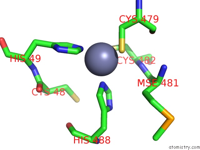
Mono view
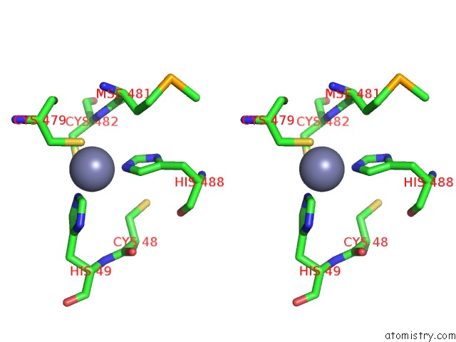
Stereo pair view
Mono view
Stereo pair view
A full contact list of Zinc with other atoms in the Zn binding
site number 1 of Crystal Structure of POZ1 and TPZ1 within 5.0Å range:
|
Zinc binding site 2 out of 2 in 5xxe
Go back to
Zinc binding site 2 out
of 2 in the Crystal Structure of POZ1 and TPZ1
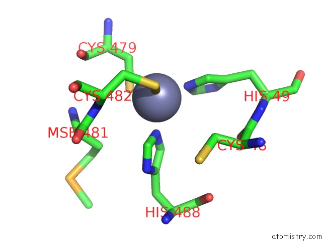
Mono view
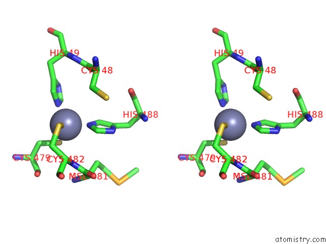
Stereo pair view
Mono view
Stereo pair view
A full contact list of Zinc with other atoms in the Zn binding
site number 2 of Crystal Structure of POZ1 and TPZ1 within 5.0Å range:
|
Reference:
J.Xue,
H.Chen,
J.Wu,
M.Takeuchi,
H.Inoue,
Y.Liu,
H.Sun,
Y.Chen,
J.Kanoh,
M.Lei.
Structure of the Fission Yeast S. Pombe Telomeric TPZ1-POZ1-RAP1 Complex. Cell Res. V. 27 1503 2017.
ISSN: ISSN 1748-7838
PubMed: 29160296
DOI: 10.1038/CR.2017.145
Page generated: Mon Oct 28 15:16:24 2024
ISSN: ISSN 1748-7838
PubMed: 29160296
DOI: 10.1038/CR.2017.145
Last articles
Zn in 9JYWZn in 9IR4
Zn in 9IR3
Zn in 9GMX
Zn in 9GMW
Zn in 9JEJ
Zn in 9ERF
Zn in 9ERE
Zn in 9EGV
Zn in 9EGW