Zinc »
PDB 5wqa-5x6h »
5wuq »
Zinc in PDB 5wuq: Crystal Structure of Sigw in Complex with Its Anti-Sigma Rsiw, A Zinc Binding Form
Protein crystallography data
The structure of Crystal Structure of Sigw in Complex with Its Anti-Sigma Rsiw, A Zinc Binding Form, PDB code: 5wuq
was solved by
S.R.Devkota,
E.Kwon,
S.C.Ha,
D.Y.Kim,
with X-Ray Crystallography technique. A brief refinement statistics is given in the table below:
| Resolution Low / High (Å) | 30.00 / 2.80 |
| Space group | P 21 21 21 |
| Cell size a, b, c (Å), α, β, γ (°) | 63.462, 64.206, 138.374, 90.00, 90.00, 90.00 |
| R / Rfree (%) | 21 / 28.7 |
Zinc Binding Sites:
The binding sites of Zinc atom in the Crystal Structure of Sigw in Complex with Its Anti-Sigma Rsiw, A Zinc Binding Form
(pdb code 5wuq). This binding sites where shown within
5.0 Angstroms radius around Zinc atom.
In total 2 binding sites of Zinc where determined in the Crystal Structure of Sigw in Complex with Its Anti-Sigma Rsiw, A Zinc Binding Form, PDB code: 5wuq:
Jump to Zinc binding site number: 1; 2;
In total 2 binding sites of Zinc where determined in the Crystal Structure of Sigw in Complex with Its Anti-Sigma Rsiw, A Zinc Binding Form, PDB code: 5wuq:
Jump to Zinc binding site number: 1; 2;
Zinc binding site 1 out of 2 in 5wuq
Go back to
Zinc binding site 1 out
of 2 in the Crystal Structure of Sigw in Complex with Its Anti-Sigma Rsiw, A Zinc Binding Form
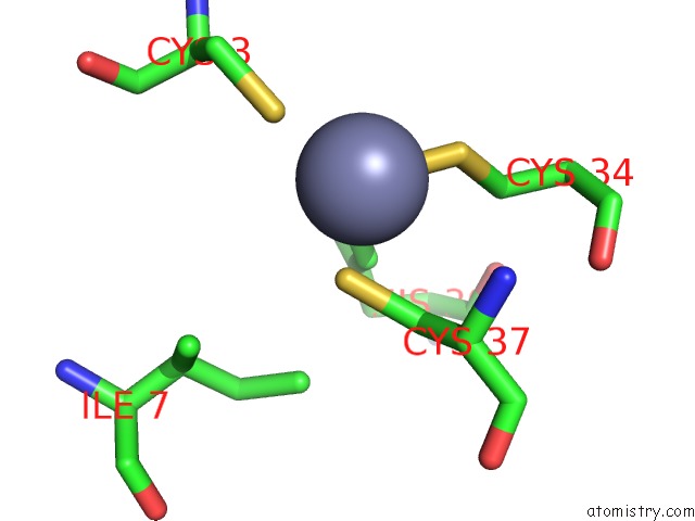
Mono view
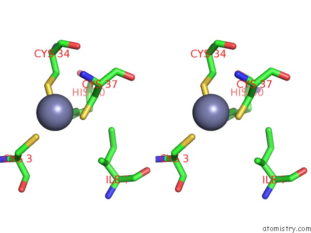
Stereo pair view
Mono view
Stereo pair view
A full contact list of Zinc with other atoms in the Zn binding
site number 1 of Crystal Structure of Sigw in Complex with Its Anti-Sigma Rsiw, A Zinc Binding Form within 5.0Å range:
|
Zinc binding site 2 out of 2 in 5wuq
Go back to
Zinc binding site 2 out
of 2 in the Crystal Structure of Sigw in Complex with Its Anti-Sigma Rsiw, A Zinc Binding Form
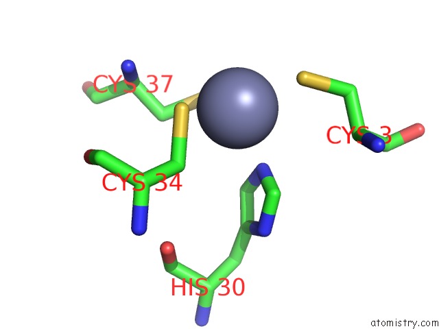
Mono view
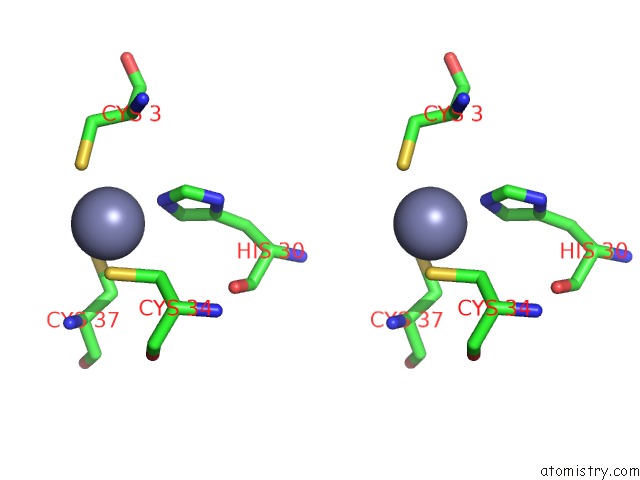
Stereo pair view
Mono view
Stereo pair view
A full contact list of Zinc with other atoms in the Zn binding
site number 2 of Crystal Structure of Sigw in Complex with Its Anti-Sigma Rsiw, A Zinc Binding Form within 5.0Å range:
|
Reference:
S.R.Devkota,
E.Kwon,
S.C.Ha,
H.W.Chang,
D.Y.Kim.
Structural Insights Into the Regulation of Bacillus Subtilis Sigw Activity By Anti-Sigma Rsiw Plos One V. 12 74284 2017.
ISSN: ESSN 1932-6203
PubMed: 28319136
DOI: 10.1371/JOURNAL.PONE.0174284
Page generated: Mon Oct 28 14:37:47 2024
ISSN: ESSN 1932-6203
PubMed: 28319136
DOI: 10.1371/JOURNAL.PONE.0174284
Last articles
Zn in 9MJ5Zn in 9HNW
Zn in 9G0L
Zn in 9FNE
Zn in 9DZN
Zn in 9E0I
Zn in 9D32
Zn in 9DAK
Zn in 8ZXC
Zn in 8ZUF