Zinc »
PDB 5vup-5w0g »
5w0g »
Zinc in PDB 5w0g: Structure of U2AF65 (U2AF2) RRM1 at 1.07 Resolution
Protein crystallography data
The structure of Structure of U2AF65 (U2AF2) RRM1 at 1.07 Resolution, PDB code: 5w0g
was solved by
A.A.Agrawal,
J.L.Jenkins,
C.L.Kielkopf,
with X-Ray Crystallography technique. A brief refinement statistics is given in the table below:
| Resolution Low / High (Å) | 16.98 / 1.07 |
| Space group | P 43 21 2 |
| Cell size a, b, c (Å), α, β, γ (°) | 28.707, 28.707, 185.851, 90.00, 90.00, 90.00 |
| R / Rfree (%) | 13.5 / 15.3 |
Zinc Binding Sites:
The binding sites of Zinc atom in the Structure of U2AF65 (U2AF2) RRM1 at 1.07 Resolution
(pdb code 5w0g). This binding sites where shown within
5.0 Angstroms radius around Zinc atom.
In total 2 binding sites of Zinc where determined in the Structure of U2AF65 (U2AF2) RRM1 at 1.07 Resolution, PDB code: 5w0g:
Jump to Zinc binding site number: 1; 2;
In total 2 binding sites of Zinc where determined in the Structure of U2AF65 (U2AF2) RRM1 at 1.07 Resolution, PDB code: 5w0g:
Jump to Zinc binding site number: 1; 2;
Zinc binding site 1 out of 2 in 5w0g
Go back to
Zinc binding site 1 out
of 2 in the Structure of U2AF65 (U2AF2) RRM1 at 1.07 Resolution
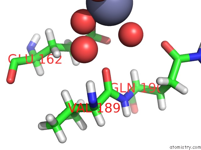
Mono view
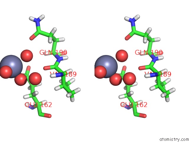
Stereo pair view
Mono view
Stereo pair view
A full contact list of Zinc with other atoms in the Zn binding
site number 1 of Structure of U2AF65 (U2AF2) RRM1 at 1.07 Resolution within 5.0Å range:
|
Zinc binding site 2 out of 2 in 5w0g
Go back to
Zinc binding site 2 out
of 2 in the Structure of U2AF65 (U2AF2) RRM1 at 1.07 Resolution
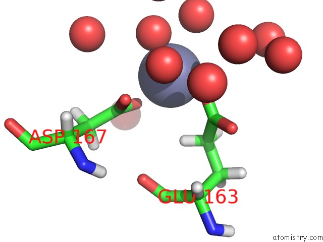
Mono view
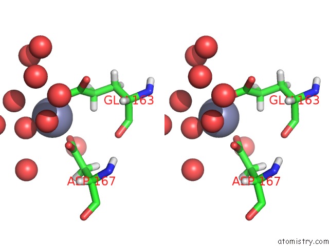
Stereo pair view
Mono view
Stereo pair view
A full contact list of Zinc with other atoms in the Zn binding
site number 2 of Structure of U2AF65 (U2AF2) RRM1 at 1.07 Resolution within 5.0Å range:
|
Reference:
E.Glasser,
A.A.Agrawal,
J.L.Jenkins,
C.L.Kielkopf.
Cancer-Associated Mutations Mapped on High-Resolution Structures of the U2AF2 Rna Recognition Motifs. Biochemistry V. 56 4757 2017.
ISSN: ISSN 1520-4995
PubMed: 28850223
DOI: 10.1021/ACS.BIOCHEM.7B00551
Page generated: Mon Oct 28 13:34:44 2024
ISSN: ISSN 1520-4995
PubMed: 28850223
DOI: 10.1021/ACS.BIOCHEM.7B00551
Last articles
Zn in 9JYWZn in 9IR4
Zn in 9IR3
Zn in 9GMX
Zn in 9GMW
Zn in 9JEJ
Zn in 9ERF
Zn in 9ERE
Zn in 9EGV
Zn in 9EGW