Zinc »
PDB 5onk-5phh »
5onk »
Zinc in PDB 5onk: Native Yndl
Protein crystallography data
The structure of Native Yndl, PDB code: 5onk
was solved by
S.Ramaswamy,
M.Rasheed,
C.Morelli,
C.Calvio,
B.Sutton,
A.Pastore,
with X-Ray Crystallography technique. A brief refinement statistics is given in the table below:
| Resolution Low / High (Å) | 34.20 / 1.03 |
| Space group | P 21 21 21 |
| Cell size a, b, c (Å), α, β, γ (°) | 38.670, 46.860, 100.070, 90.00, 90.00, 90.00 |
| R / Rfree (%) | 16.6 / 18.7 |
Zinc Binding Sites:
The binding sites of Zinc atom in the Native Yndl
(pdb code 5onk). This binding sites where shown within
5.0 Angstroms radius around Zinc atom.
In total only one binding site of Zinc was determined in the Native Yndl, PDB code: 5onk:
In total only one binding site of Zinc was determined in the Native Yndl, PDB code: 5onk:
Zinc binding site 1 out of 1 in 5onk
Go back to
Zinc binding site 1 out
of 1 in the Native Yndl

Mono view
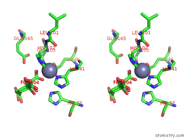
Stereo pair view
Mono view
Stereo pair view
A full contact list of Zinc with other atoms in the Zn binding
site number 1 of Native Yndl within 5.0Å range:
|
Reference:
S.Ramaswamy,
M.Rasheed,
C.F.Morelli,
C.Calvio,
B.J.Sutton,
A.Pastore.
The Structure of Pghl Hydrolase Bound to Its Substrate Poly-Gamma-Glutamate. Febs J. V. 285 4575 2018.
ISSN: ISSN 1742-4658
PubMed: 30387270
DOI: 10.1111/FEBS.14688
Page generated: Sun Oct 27 23:44:47 2024
ISSN: ISSN 1742-4658
PubMed: 30387270
DOI: 10.1111/FEBS.14688
Last articles
Zn in 9MJ5Zn in 9HNW
Zn in 9G0L
Zn in 9FNE
Zn in 9DZN
Zn in 9E0I
Zn in 9D32
Zn in 9DAK
Zn in 8ZXC
Zn in 8ZUF