Zinc »
PDB 5msb-5n2x »
5n0b »
Zinc in PDB 5n0b: Crystal Structure of the Tetanus Neurotoxin in Complex with GD1A
Enzymatic activity of Crystal Structure of the Tetanus Neurotoxin in Complex with GD1A
All present enzymatic activity of Crystal Structure of the Tetanus Neurotoxin in Complex with GD1A:
3.4.24.68;
3.4.24.68;
Protein crystallography data
The structure of Crystal Structure of the Tetanus Neurotoxin in Complex with GD1A, PDB code: 5n0b
was solved by
G.Masuyer,
J.Conrad,
P.Stenmark,
with X-Ray Crystallography technique. A brief refinement statistics is given in the table below:
| Resolution Low / High (Å) | 68.50 / 2.60 |
| Space group | C 1 2 1 |
| Cell size a, b, c (Å), α, β, γ (°) | 152.695, 136.840, 92.630, 90.00, 90.07, 90.00 |
| R / Rfree (%) | 20.3 / 24.7 |
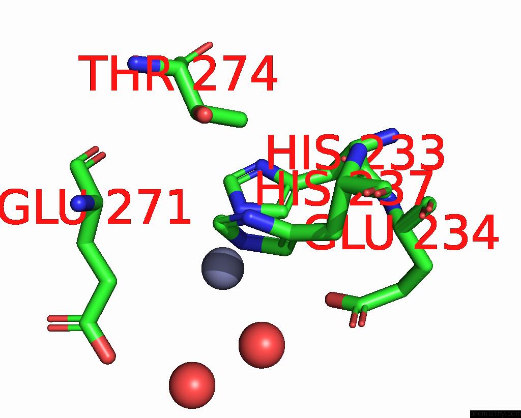
Zinc Binding Sites:
The binding sites of Zinc atom in the Crystal Structure of the Tetanus Neurotoxin in Complex with GD1A
(pdb code 5n0b). This binding sites where shown within
5.0 Angstroms radius around Zinc atom.
In total only one binding site of Zinc was determined in the Crystal Structure of the Tetanus Neurotoxin in Complex with GD1A, PDB code: 5n0b:
In total only one binding site of Zinc was determined in the Crystal Structure of the Tetanus Neurotoxin in Complex with GD1A, PDB code: 5n0b:
Zinc binding site 1 out of 1 in 5n0b
Go back to
Zinc binding site 1 out
of 1 in the Crystal Structure of the Tetanus Neurotoxin in Complex with GD1A
Mono view

Stereo pair view
Mono view
Stereo pair view
A full contact list of Zinc with other atoms in the Zn binding
site number 1 of Crystal Structure of the Tetanus Neurotoxin in Complex with GD1A within 5.0Å range:
|
Reference:
G.Masuyer,
J.Conrad,
P.Stenmark.
The Structure of the Tetanus Toxin Reveals pH-Mediated Domain Dynamics. Embo Rep. V. 18 1306 2017.
ISSN: ESSN 1469-3178
PubMed: 28645943
DOI: 10.15252/EMBR.201744198
Page generated: Sun Oct 27 22:25:39 2024
ISSN: ESSN 1469-3178
PubMed: 28645943
DOI: 10.15252/EMBR.201744198
Last articles
Zn in 9JYWZn in 9IR4
Zn in 9IR3
Zn in 9GMX
Zn in 9GMW
Zn in 9JEJ
Zn in 9ERF
Zn in 9ERE
Zn in 9EGV
Zn in 9EGW