Zinc »
PDB 5msb-5n2x »
5mte »
Zinc in PDB 5mte: Crystal Structure of Pdf From the Vibrio Parahaemolyticus Bacteriophage VP16T in Complex with Actinonin - Crystal Form II
Protein crystallography data
The structure of Crystal Structure of Pdf From the Vibrio Parahaemolyticus Bacteriophage VP16T in Complex with Actinonin - Crystal Form II, PDB code: 5mte
was solved by
S.Fieulaine,
R.Grzela,
C.Giglione,
T.Meinnel,
with X-Ray Crystallography technique. A brief refinement statistics is given in the table below:
| Resolution Low / High (Å) | 41.67 / 1.40 |
| Space group | P 32 2 1 |
| Cell size a, b, c (Å), α, β, γ (°) | 64.620, 64.620, 124.890, 90.00, 90.00, 120.00 |
| R / Rfree (%) | 18.7 / 20.3 |
Other elements in 5mte:
The structure of Crystal Structure of Pdf From the Vibrio Parahaemolyticus Bacteriophage VP16T in Complex with Actinonin - Crystal Form II also contains other interesting chemical elements:
| Nickel | (Ni) | 3 atoms |
Zinc Binding Sites:
The binding sites of Zinc atom in the Crystal Structure of Pdf From the Vibrio Parahaemolyticus Bacteriophage VP16T in Complex with Actinonin - Crystal Form II
(pdb code 5mte). This binding sites where shown within
5.0 Angstroms radius around Zinc atom.
In total 2 binding sites of Zinc where determined in the Crystal Structure of Pdf From the Vibrio Parahaemolyticus Bacteriophage VP16T in Complex with Actinonin - Crystal Form II, PDB code: 5mte:
Jump to Zinc binding site number: 1; 2;
In total 2 binding sites of Zinc where determined in the Crystal Structure of Pdf From the Vibrio Parahaemolyticus Bacteriophage VP16T in Complex with Actinonin - Crystal Form II, PDB code: 5mte:
Jump to Zinc binding site number: 1; 2;
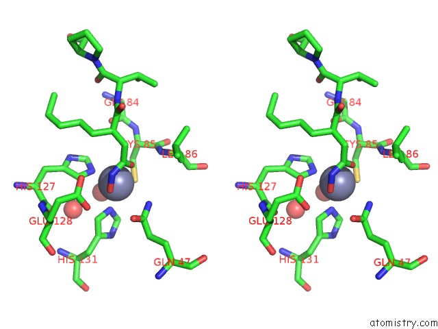
Zinc binding site 1 out of 2 in 5mte
Go back to
Zinc binding site 1 out
of 2 in the Crystal Structure of Pdf From the Vibrio Parahaemolyticus Bacteriophage VP16T in Complex with Actinonin - Crystal Form II
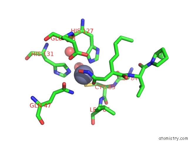
Mono view
Stereo pair view
Mono view
Stereo pair view
A full contact list of Zinc with other atoms in the Zn binding
site number 1 of Crystal Structure of Pdf From the Vibrio Parahaemolyticus Bacteriophage VP16T in Complex with Actinonin - Crystal Form II within 5.0Å range:
|
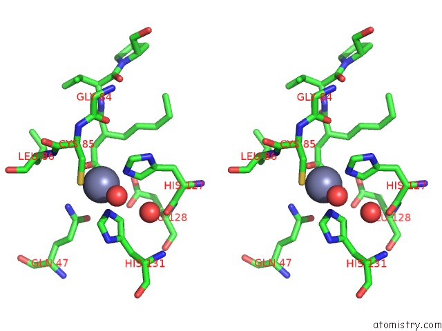
Zinc binding site 2 out of 2 in 5mte
Go back to
Zinc binding site 2 out
of 2 in the Crystal Structure of Pdf From the Vibrio Parahaemolyticus Bacteriophage VP16T in Complex with Actinonin - Crystal Form II
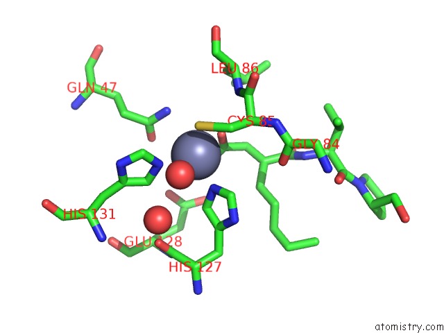
Mono view
Stereo pair view
Mono view
Stereo pair view
A full contact list of Zinc with other atoms in the Zn binding
site number 2 of Crystal Structure of Pdf From the Vibrio Parahaemolyticus Bacteriophage VP16T in Complex with Actinonin - Crystal Form II within 5.0Å range:
|
Reference:
R.Grzela,
J.Nusbaum,
S.Fieulaine,
F.Lavecchia,
M.Desmadril,
N.Nhiri,
A.Van Dorsselaer,
S.Cianferani,
E.Jacquet,
T.Meinnel,
C.Giglione.
Peptide Deformylases From Vibrio Parahaemolyticus Phage and Bacteria Display Similar Deformylase Activity and Inhibitor Binding Clefts. Biochim. Biophys. Acta V.1866 348 2018.
ISSN: ISSN 0006-3002
PubMed: 29101077
DOI: 10.1016/J.BBAPAP.2017.10.007
Page generated: Sun Oct 27 22:20:12 2024
ISSN: ISSN 0006-3002
PubMed: 29101077
DOI: 10.1016/J.BBAPAP.2017.10.007
Last articles
Zn in 9JYWZn in 9IR4
Zn in 9IR3
Zn in 9GMX
Zn in 9GMW
Zn in 9JEJ
Zn in 9ERF
Zn in 9ERE
Zn in 9EGV
Zn in 9EGW