Zinc »
PDB 5i3b-5ijq »
5ib9 »
Zinc in PDB 5ib9: Crystal Structure of Aminopeptidase Equipped with Pad From Aneurinibacillus Sp. Am-1
Protein crystallography data
The structure of Crystal Structure of Aminopeptidase Equipped with Pad From Aneurinibacillus Sp. Am-1, PDB code: 5ib9
was solved by
R.Tagawa,
H.Nakano,
K.Watanabe,
with X-Ray Crystallography technique. A brief refinement statistics is given in the table below:
| Resolution Low / High (Å) | 39.80 / 1.40 |
| Space group | P 21 21 2 |
| Cell size a, b, c (Å), α, β, γ (°) | 93.180, 68.443, 76.589, 90.00, 90.00, 90.00 |
| R / Rfree (%) | 17.9 / 22.3 |
Zinc Binding Sites:
The binding sites of Zinc atom in the Crystal Structure of Aminopeptidase Equipped with Pad From Aneurinibacillus Sp. Am-1
(pdb code 5ib9). This binding sites where shown within
5.0 Angstroms radius around Zinc atom.
In total 6 binding sites of Zinc where determined in the Crystal Structure of Aminopeptidase Equipped with Pad From Aneurinibacillus Sp. Am-1, PDB code: 5ib9:
Jump to Zinc binding site number: 1; 2; 3; 4; 5; 6;
In total 6 binding sites of Zinc where determined in the Crystal Structure of Aminopeptidase Equipped with Pad From Aneurinibacillus Sp. Am-1, PDB code: 5ib9:
Jump to Zinc binding site number: 1; 2; 3; 4; 5; 6;
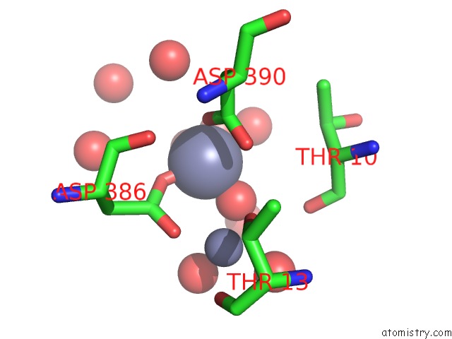
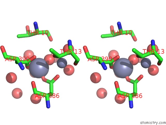
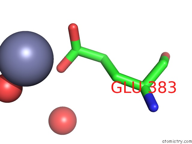
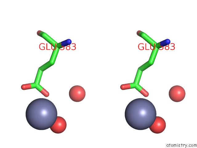
Zinc binding site 1 out of 6 in 5ib9
Go back to
Zinc binding site 1 out
of 6 in the Crystal Structure of Aminopeptidase Equipped with Pad From Aneurinibacillus Sp. Am-1
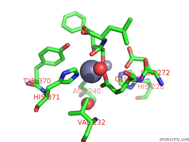
Mono view
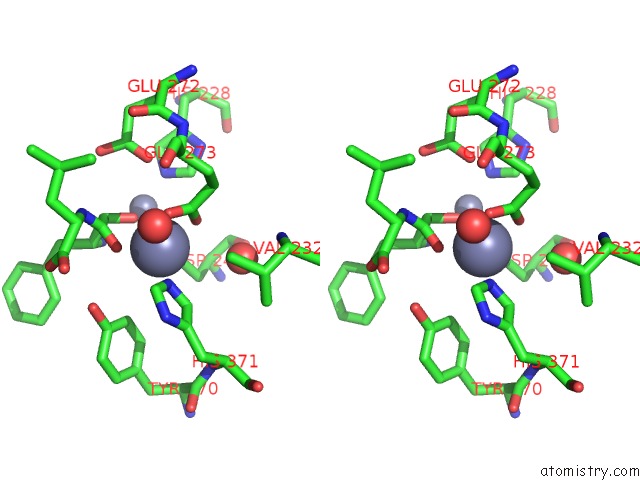
Stereo pair view
Mono view
Stereo pair view
A full contact list of Zinc with other atoms in the Zn binding
site number 1 of Crystal Structure of Aminopeptidase Equipped with Pad From Aneurinibacillus Sp. Am-1 within 5.0Å range:
|
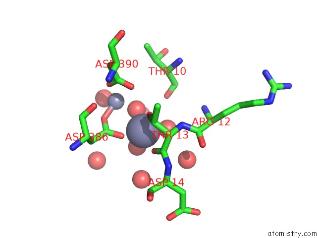
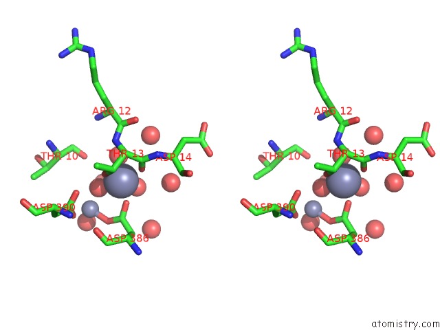
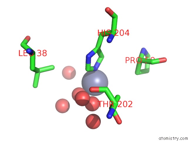
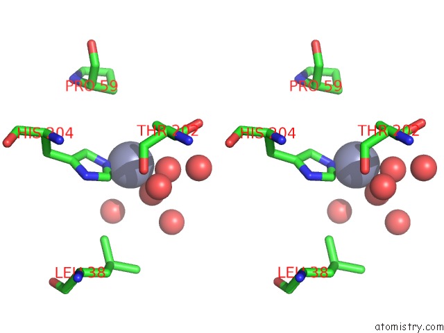
Zinc binding site 2 out of 6 in 5ib9
Go back to
Zinc binding site 2 out
of 6 in the Crystal Structure of Aminopeptidase Equipped with Pad From Aneurinibacillus Sp. Am-1
Mono view
Stereo pair view
Mono view
Stereo pair view
A full contact list of Zinc with other atoms in the Zn binding
site number 2 of Crystal Structure of Aminopeptidase Equipped with Pad From Aneurinibacillus Sp. Am-1 within 5.0Å range:
|
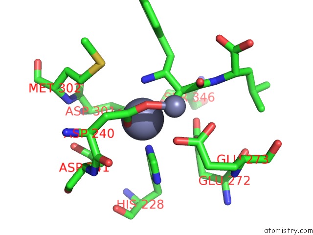
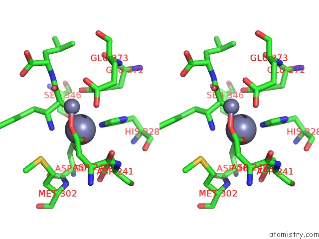
Zinc binding site 3 out of 6 in 5ib9
Go back to
Zinc binding site 3 out
of 6 in the Crystal Structure of Aminopeptidase Equipped with Pad From Aneurinibacillus Sp. Am-1
Mono view
Stereo pair view
Mono view
Stereo pair view
A full contact list of Zinc with other atoms in the Zn binding
site number 3 of Crystal Structure of Aminopeptidase Equipped with Pad From Aneurinibacillus Sp. Am-1 within 5.0Å range:
|
Zinc binding site 4 out of 6 in 5ib9
Go back to
Zinc binding site 4 out
of 6 in the Crystal Structure of Aminopeptidase Equipped with Pad From Aneurinibacillus Sp. Am-1
Mono view
Stereo pair view
Mono view
Stereo pair view
A full contact list of Zinc with other atoms in the Zn binding
site number 4 of Crystal Structure of Aminopeptidase Equipped with Pad From Aneurinibacillus Sp. Am-1 within 5.0Å range:
|
Zinc binding site 5 out of 6 in 5ib9
Go back to
Zinc binding site 5 out
of 6 in the Crystal Structure of Aminopeptidase Equipped with Pad From Aneurinibacillus Sp. Am-1
Mono view
Stereo pair view
Mono view
Stereo pair view
A full contact list of Zinc with other atoms in the Zn binding
site number 5 of Crystal Structure of Aminopeptidase Equipped with Pad From Aneurinibacillus Sp. Am-1 within 5.0Å range:
|
Zinc binding site 6 out of 6 in 5ib9
Go back to
Zinc binding site 6 out
of 6 in the Crystal Structure of Aminopeptidase Equipped with Pad From Aneurinibacillus Sp. Am-1
Mono view
Stereo pair view
Mono view
Stereo pair view
A full contact list of Zinc with other atoms in the Zn binding
site number 6 of Crystal Structure of Aminopeptidase Equipped with Pad From Aneurinibacillus Sp. Am-1 within 5.0Å range:
|
Reference:
R.Tagawa,
H.Nakano,
K.Watanabe.
Crystal Structure of Aminopeptidase To Be Published.
Page generated: Sun Oct 27 18:01:52 2024
Last articles
Zn in 9JYWZn in 9IR4
Zn in 9IR3
Zn in 9GMX
Zn in 9GMW
Zn in 9JEJ
Zn in 9ERF
Zn in 9ERE
Zn in 9EGV
Zn in 9EGW