Zinc »
PDB 5fpb-5fvt »
5fth »
Zinc in PDB 5fth: Crystal Structure of the GLUA2 K738M-T744K Lbd in Complex with Glutamate (Zinc Form)
Protein crystallography data
The structure of Crystal Structure of the GLUA2 K738M-T744K Lbd in Complex with Glutamate (Zinc Form), PDB code: 5fth
was solved by
N.Nayeem,
T.Green,
with X-Ray Crystallography technique. A brief refinement statistics is given in the table below:
| Resolution Low / High (Å) | 92.208 / 2.90 |
| Space group | P 2 21 21 |
| Cell size a, b, c (Å), α, β, γ (°) | 46.375, 110.523, 167.245, 90.00, 90.00, 90.00 |
| R / Rfree (%) | 24.29 / 28.29 |
Zinc Binding Sites:
The binding sites of Zinc atom in the Crystal Structure of the GLUA2 K738M-T744K Lbd in Complex with Glutamate (Zinc Form)
(pdb code 5fth). This binding sites where shown within
5.0 Angstroms radius around Zinc atom.
In total 5 binding sites of Zinc where determined in the Crystal Structure of the GLUA2 K738M-T744K Lbd in Complex with Glutamate (Zinc Form), PDB code: 5fth:
Jump to Zinc binding site number: 1; 2; 3; 4; 5;
In total 5 binding sites of Zinc where determined in the Crystal Structure of the GLUA2 K738M-T744K Lbd in Complex with Glutamate (Zinc Form), PDB code: 5fth:
Jump to Zinc binding site number: 1; 2; 3; 4; 5;
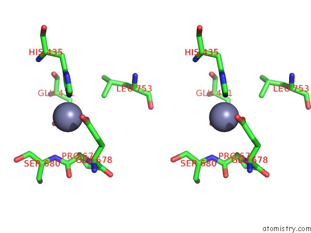
Zinc binding site 1 out of 5 in 5fth
Go back to
Zinc binding site 1 out
of 5 in the Crystal Structure of the GLUA2 K738M-T744K Lbd in Complex with Glutamate (Zinc Form)

Mono view

Stereo pair view
Mono view
Stereo pair view
A full contact list of Zinc with other atoms in the Zn binding
site number 1 of Crystal Structure of the GLUA2 K738M-T744K Lbd in Complex with Glutamate (Zinc Form) within 5.0Å range:
|
Zinc binding site 2 out of 5 in 5fth
Go back to
Zinc binding site 2 out
of 5 in the Crystal Structure of the GLUA2 K738M-T744K Lbd in Complex with Glutamate (Zinc Form)

Mono view
Stereo pair view
Mono view
Stereo pair view
A full contact list of Zinc with other atoms in the Zn binding
site number 2 of Crystal Structure of the GLUA2 K738M-T744K Lbd in Complex with Glutamate (Zinc Form) within 5.0Å range:
|
Zinc binding site 3 out of 5 in 5fth
Go back to
Zinc binding site 3 out
of 5 in the Crystal Structure of the GLUA2 K738M-T744K Lbd in Complex with Glutamate (Zinc Form)

Mono view

Stereo pair view
Mono view
Stereo pair view
A full contact list of Zinc with other atoms in the Zn binding
site number 3 of Crystal Structure of the GLUA2 K738M-T744K Lbd in Complex with Glutamate (Zinc Form) within 5.0Å range:
|
Zinc binding site 4 out of 5 in 5fth
Go back to
Zinc binding site 4 out
of 5 in the Crystal Structure of the GLUA2 K738M-T744K Lbd in Complex with Glutamate (Zinc Form)

Mono view

Stereo pair view
Mono view
Stereo pair view
A full contact list of Zinc with other atoms in the Zn binding
site number 4 of Crystal Structure of the GLUA2 K738M-T744K Lbd in Complex with Glutamate (Zinc Form) within 5.0Å range:
|
Zinc binding site 5 out of 5 in 5fth
Go back to
Zinc binding site 5 out
of 5 in the Crystal Structure of the GLUA2 K738M-T744K Lbd in Complex with Glutamate (Zinc Form)

Mono view

Stereo pair view
Mono view
Stereo pair view
A full contact list of Zinc with other atoms in the Zn binding
site number 5 of Crystal Structure of the GLUA2 K738M-T744K Lbd in Complex with Glutamate (Zinc Form) within 5.0Å range:
|
Reference:
G.B.Dawe,
M.Musgaard,
M.R.P.Aurousseau,
N.Nayeem,
T.Green,
P.C.Biggin,
D.Bowie.
Distinct Structural Pathways Coordinate the Activation of Ampa Receptor-Auxiliary Subunit Complexes. Neuron V. 89 1264 2016.
ISSN: ISSN 0896-6273
PubMed: 26924438
DOI: 10.1016/J.NEURON.2016.01.038
Page generated: Sun Oct 27 16:30:08 2024
ISSN: ISSN 0896-6273
PubMed: 26924438
DOI: 10.1016/J.NEURON.2016.01.038
Last articles
Zn in 9JYWZn in 9IR4
Zn in 9IR3
Zn in 9GMX
Zn in 9GMW
Zn in 9JEJ
Zn in 9ERF
Zn in 9ERE
Zn in 9EGV
Zn in 9EGW