Zinc »
PDB 5f8h-5fi3 »
5fch »
Zinc in PDB 5fch: Crystal Structure of Xaa-Pro Dipeptidase From Xanthomonas Campestris, Phosphate and Zn Bound
Enzymatic activity of Crystal Structure of Xaa-Pro Dipeptidase From Xanthomonas Campestris, Phosphate and Zn Bound
All present enzymatic activity of Crystal Structure of Xaa-Pro Dipeptidase From Xanthomonas Campestris, Phosphate and Zn Bound:
3.4.13.9;
3.4.13.9;
Protein crystallography data
The structure of Crystal Structure of Xaa-Pro Dipeptidase From Xanthomonas Campestris, Phosphate and Zn Bound, PDB code: 5fch
was solved by
A.Kumar,
V.Are,
B.Ghosh,
S.Jamdar,
R.D.Makde,
with X-Ray Crystallography technique. A brief refinement statistics is given in the table below:
| Resolution Low / High (Å) | 45.00 / 1.95 |
| Space group | P 21 21 21 |
| Cell size a, b, c (Å), α, β, γ (°) | 82.032, 104.080, 112.412, 90.00, 90.00, 90.00 |
| R / Rfree (%) | 17.2 / 21.1 |
Zinc Binding Sites:
The binding sites of Zinc atom in the Crystal Structure of Xaa-Pro Dipeptidase From Xanthomonas Campestris, Phosphate and Zn Bound
(pdb code 5fch). This binding sites where shown within
5.0 Angstroms radius around Zinc atom.
In total 4 binding sites of Zinc where determined in the Crystal Structure of Xaa-Pro Dipeptidase From Xanthomonas Campestris, Phosphate and Zn Bound, PDB code: 5fch:
Jump to Zinc binding site number: 1; 2; 3; 4;
In total 4 binding sites of Zinc where determined in the Crystal Structure of Xaa-Pro Dipeptidase From Xanthomonas Campestris, Phosphate and Zn Bound, PDB code: 5fch:
Jump to Zinc binding site number: 1; 2; 3; 4;
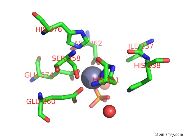
Zinc binding site 1 out of 4 in 5fch
Go back to
Zinc binding site 1 out
of 4 in the Crystal Structure of Xaa-Pro Dipeptidase From Xanthomonas Campestris, Phosphate and Zn Bound
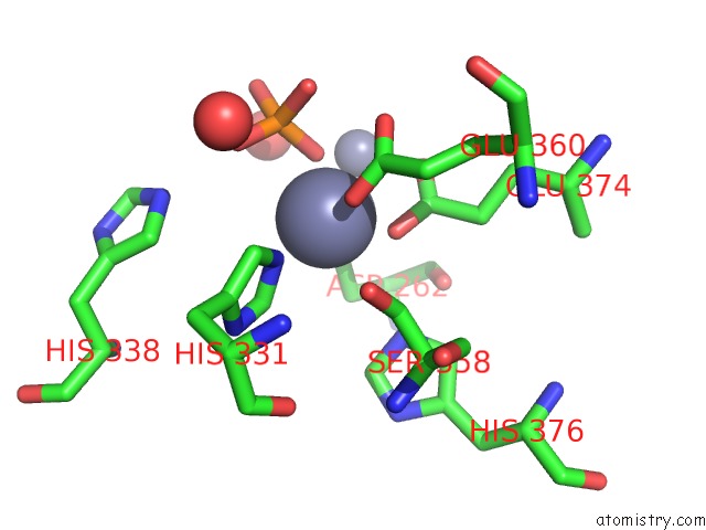
Mono view
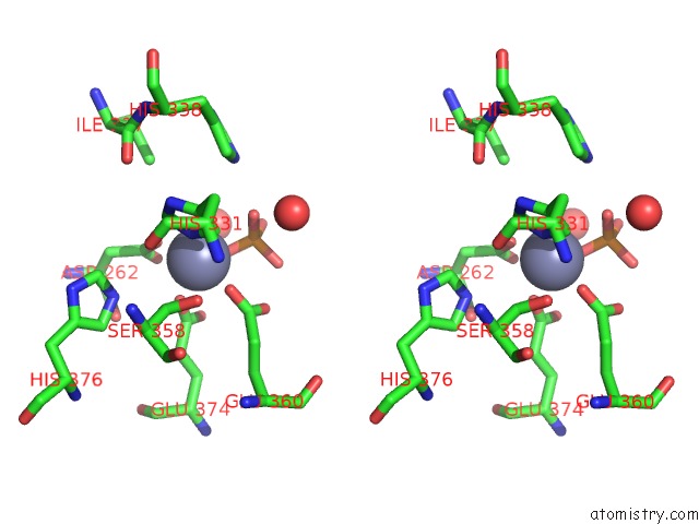
Stereo pair view
Mono view
Stereo pair view
A full contact list of Zinc with other atoms in the Zn binding
site number 1 of Crystal Structure of Xaa-Pro Dipeptidase From Xanthomonas Campestris, Phosphate and Zn Bound within 5.0Å range:
|
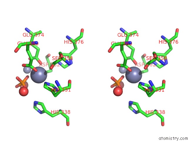
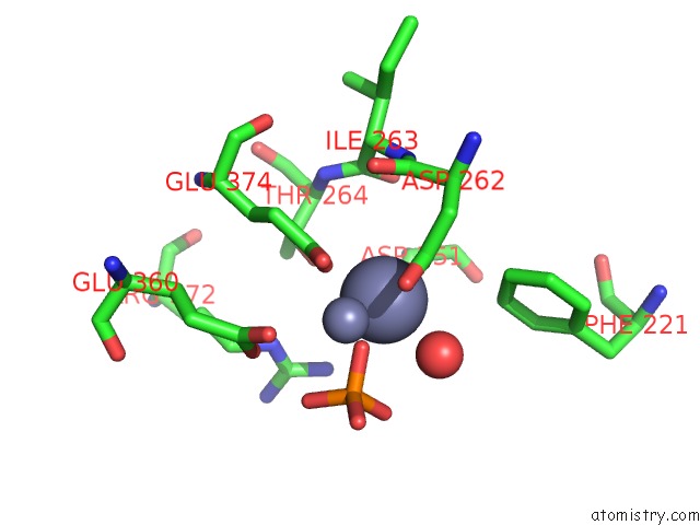
Zinc binding site 2 out of 4 in 5fch
Go back to
Zinc binding site 2 out
of 4 in the Crystal Structure of Xaa-Pro Dipeptidase From Xanthomonas Campestris, Phosphate and Zn Bound
Mono view
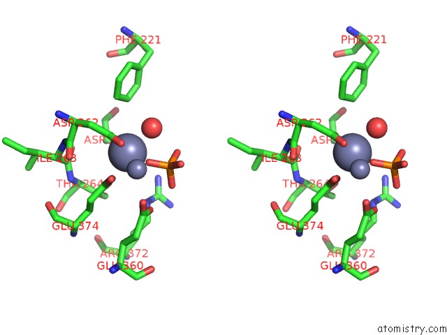
Stereo pair view
Mono view
Stereo pair view
A full contact list of Zinc with other atoms in the Zn binding
site number 2 of Crystal Structure of Xaa-Pro Dipeptidase From Xanthomonas Campestris, Phosphate and Zn Bound within 5.0Å range:
|
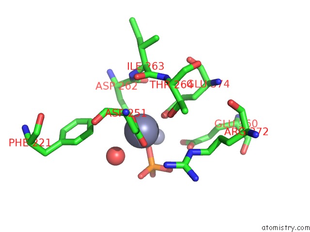
Zinc binding site 3 out of 4 in 5fch
Go back to
Zinc binding site 3 out
of 4 in the Crystal Structure of Xaa-Pro Dipeptidase From Xanthomonas Campestris, Phosphate and Zn Bound
Mono view
Stereo pair view
Mono view
Stereo pair view
A full contact list of Zinc with other atoms in the Zn binding
site number 3 of Crystal Structure of Xaa-Pro Dipeptidase From Xanthomonas Campestris, Phosphate and Zn Bound within 5.0Å range:
|
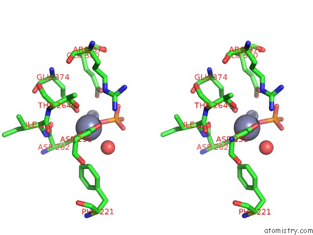
Zinc binding site 4 out of 4 in 5fch
Go back to
Zinc binding site 4 out
of 4 in the Crystal Structure of Xaa-Pro Dipeptidase From Xanthomonas Campestris, Phosphate and Zn Bound
Mono view
Stereo pair view
Mono view
Stereo pair view
A full contact list of Zinc with other atoms in the Zn binding
site number 4 of Crystal Structure of Xaa-Pro Dipeptidase From Xanthomonas Campestris, Phosphate and Zn Bound within 5.0Å range:
|
Reference:
V.N.Are,
A.Kumar,
S.Kumar,
V.D.Goyal,
B.Ghosh,
D.Bhatnagar,
S.N.Jamdar,
R.D.Makde.
Crystal Structure and Biochemical Investigations Reveal Novel Mode of Substrate Selectivity and Illuminate Substrate Inhibition and Allostericity in A Subfamily of Xaa-Pro Dipeptidases Biochim. Biophys. Acta V.1865 153 2017.
ISSN: ISSN 0006-3002
PubMed: 27816563
DOI: 10.1016/J.BBAPAP.2016.10.016
Page generated: Sun Oct 27 16:02:42 2024
ISSN: ISSN 0006-3002
PubMed: 27816563
DOI: 10.1016/J.BBAPAP.2016.10.016
Last articles
Zn in 9JYWZn in 9IR4
Zn in 9IR3
Zn in 9GMX
Zn in 9GMW
Zn in 9JEJ
Zn in 9ERF
Zn in 9ERE
Zn in 9EGV
Zn in 9EGW