Zinc »
PDB 5e8c-5ehe »
5ecg »
Zinc in PDB 5ecg: Crystal Structure of the Brct Domains of 53BP1 in Complex with P53 and H2AX-PSER139 (GAMMAH2AX)
Protein crystallography data
The structure of Crystal Structure of the Brct Domains of 53BP1 in Complex with P53 and H2AX-PSER139 (GAMMAH2AX), PDB code: 5ecg
was solved by
M.Day,
A.W.Oliver,
L.H.Pearl,
with X-Ray Crystallography technique. A brief refinement statistics is given in the table below:
| Resolution Low / High (Å) | 48.06 / 3.00 |
| Space group | P 21 21 21 |
| Cell size a, b, c (Å), α, β, γ (°) | 70.264, 94.498, 131.742, 90.00, 90.00, 90.00 |
| R / Rfree (%) | 20.4 / 26.4 |
Zinc Binding Sites:
The binding sites of Zinc atom in the Crystal Structure of the Brct Domains of 53BP1 in Complex with P53 and H2AX-PSER139 (GAMMAH2AX)
(pdb code 5ecg). This binding sites where shown within
5.0 Angstroms radius around Zinc atom.
In total 2 binding sites of Zinc where determined in the Crystal Structure of the Brct Domains of 53BP1 in Complex with P53 and H2AX-PSER139 (GAMMAH2AX), PDB code: 5ecg:
Jump to Zinc binding site number: 1; 2;
In total 2 binding sites of Zinc where determined in the Crystal Structure of the Brct Domains of 53BP1 in Complex with P53 and H2AX-PSER139 (GAMMAH2AX), PDB code: 5ecg:
Jump to Zinc binding site number: 1; 2;
Zinc binding site 1 out of 2 in 5ecg
Go back to
Zinc binding site 1 out
of 2 in the Crystal Structure of the Brct Domains of 53BP1 in Complex with P53 and H2AX-PSER139 (GAMMAH2AX)
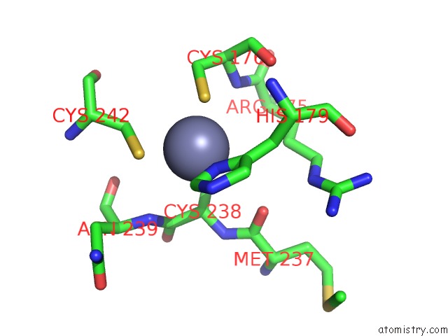
Mono view
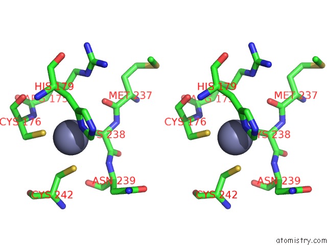
Stereo pair view
Mono view
Stereo pair view
A full contact list of Zinc with other atoms in the Zn binding
site number 1 of Crystal Structure of the Brct Domains of 53BP1 in Complex with P53 and H2AX-PSER139 (GAMMAH2AX) within 5.0Å range:
|
Zinc binding site 2 out of 2 in 5ecg
Go back to
Zinc binding site 2 out
of 2 in the Crystal Structure of the Brct Domains of 53BP1 in Complex with P53 and H2AX-PSER139 (GAMMAH2AX)
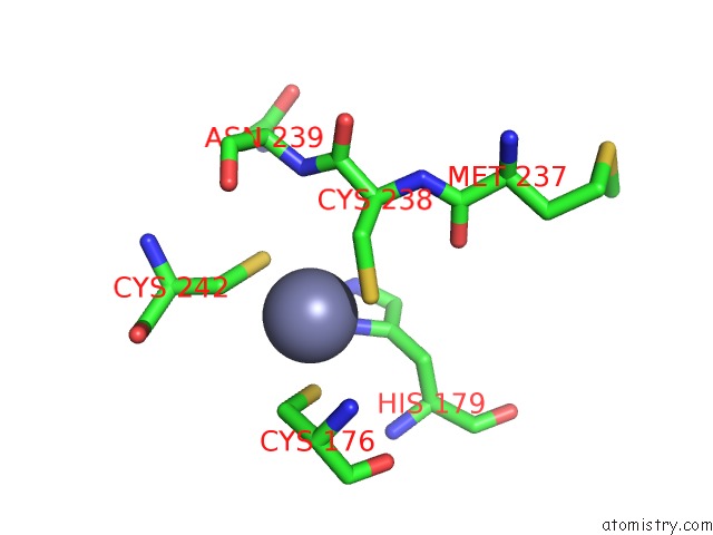
Mono view
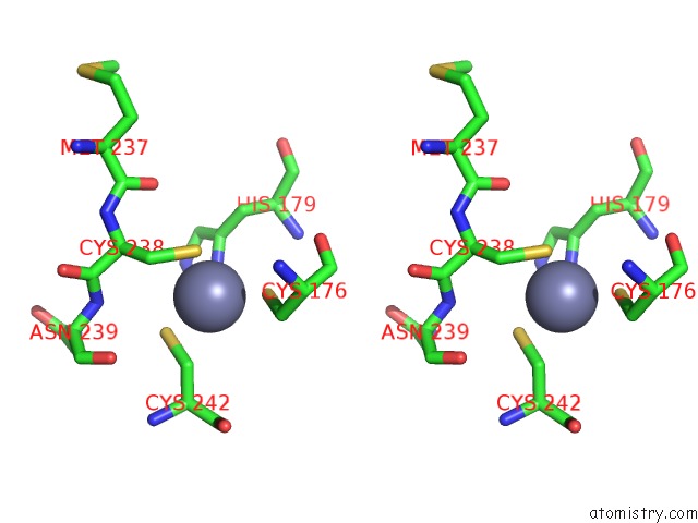
Stereo pair view
Mono view
Stereo pair view
A full contact list of Zinc with other atoms in the Zn binding
site number 2 of Crystal Structure of the Brct Domains of 53BP1 in Complex with P53 and H2AX-PSER139 (GAMMAH2AX) within 5.0Å range:
|
Reference:
R.A.Baldock,
M.Day,
O.J.Wilkinson,
R.Cloney,
P.A.Jeggo,
A.W.Oliver,
F.Z.Watts,
L.H.Pearl.
Atm Localization and Heterochromatin Repair Depend on Direct Interaction of the 53BP1-BRCT2 Domain with Gamma H2AX. Cell Rep V. 13 2081 2015.
ISSN: ESSN 2211-1247
PubMed: 26628370
DOI: 10.1016/J.CELREP.2015.10.074
Page generated: Sun Oct 27 15:06:51 2024
ISSN: ESSN 2211-1247
PubMed: 26628370
DOI: 10.1016/J.CELREP.2015.10.074
Last articles
Zn in 9JYWZn in 9IR4
Zn in 9IR3
Zn in 9GMX
Zn in 9GMW
Zn in 9JEJ
Zn in 9ERF
Zn in 9ERE
Zn in 9EGV
Zn in 9EGW