Zinc »
PDB 5amj-5b2p »
5b16 »
Zinc in PDB 5b16: X-Ray Structure of Drosha in Complex with the C-Terminal Tail of DGCR8.
Enzymatic activity of X-Ray Structure of Drosha in Complex with the C-Terminal Tail of DGCR8.
All present enzymatic activity of X-Ray Structure of Drosha in Complex with the C-Terminal Tail of DGCR8.:
3.1.26.3;
3.1.26.3;
Protein crystallography data
The structure of X-Ray Structure of Drosha in Complex with the C-Terminal Tail of DGCR8., PDB code: 5b16
was solved by
S.C.Kwon,
T.A.Nguyen,
Y.G.Choi,
M.H.Jo,
S.Hohng,
V.N.Kim,
J.S.Woo,
with X-Ray Crystallography technique. A brief refinement statistics is given in the table below:
| Resolution Low / High (Å) | 19.93 / 3.20 |
| Space group | C 1 2 1 |
| Cell size a, b, c (Å), α, β, γ (°) | 117.254, 118.136, 122.296, 90.00, 102.07, 90.00 |
| R / Rfree (%) | 26.7 / 30 |
Zinc Binding Sites:
The binding sites of Zinc atom in the X-Ray Structure of Drosha in Complex with the C-Terminal Tail of DGCR8.
(pdb code 5b16). This binding sites where shown within
5.0 Angstroms radius around Zinc atom.
In total 2 binding sites of Zinc where determined in the X-Ray Structure of Drosha in Complex with the C-Terminal Tail of DGCR8., PDB code: 5b16:
Jump to Zinc binding site number: 1; 2;
In total 2 binding sites of Zinc where determined in the X-Ray Structure of Drosha in Complex with the C-Terminal Tail of DGCR8., PDB code: 5b16:
Jump to Zinc binding site number: 1; 2;
Zinc binding site 1 out of 2 in 5b16
Go back to
Zinc binding site 1 out
of 2 in the X-Ray Structure of Drosha in Complex with the C-Terminal Tail of DGCR8.
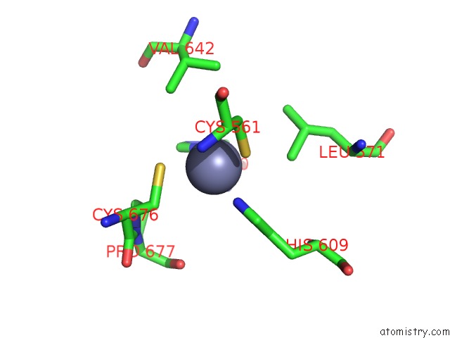
Mono view
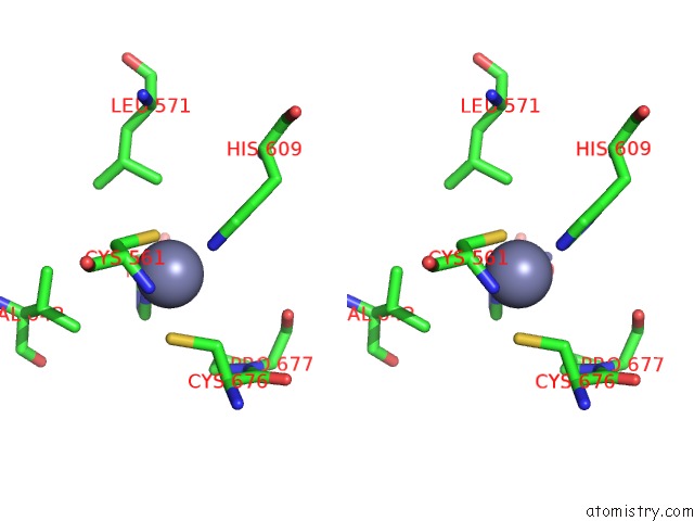
Stereo pair view
Mono view
Stereo pair view
A full contact list of Zinc with other atoms in the Zn binding
site number 1 of X-Ray Structure of Drosha in Complex with the C-Terminal Tail of DGCR8. within 5.0Å range:
|
Zinc binding site 2 out of 2 in 5b16
Go back to
Zinc binding site 2 out
of 2 in the X-Ray Structure of Drosha in Complex with the C-Terminal Tail of DGCR8.
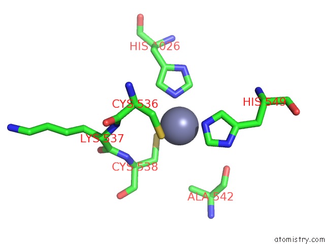
Mono view
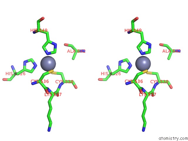
Stereo pair view
Mono view
Stereo pair view
A full contact list of Zinc with other atoms in the Zn binding
site number 2 of X-Ray Structure of Drosha in Complex with the C-Terminal Tail of DGCR8. within 5.0Å range:
|
Reference:
S.C.Kwon,
T.A.Nguyen,
Y.G.Choi,
M.H.Jo,
S.Hohng,
V.N.Kim,
J.S.Woo.
Structure of Human Drosha Cell V. 164 81 2016.
ISSN: ISSN 1097-4172
PubMed: 26748718
DOI: 10.1016/J.CELL.2015.12.019
Page generated: Sun Oct 27 13:17:29 2024
ISSN: ISSN 1097-4172
PubMed: 26748718
DOI: 10.1016/J.CELL.2015.12.019
Last articles
Zn in 9JYWZn in 9IR4
Zn in 9IR3
Zn in 9GMX
Zn in 9GMW
Zn in 9JEJ
Zn in 9ERF
Zn in 9ERE
Zn in 9EGV
Zn in 9EGW