Zinc »
PDB 5amj-5b2p »
5b0h »
Zinc in PDB 5b0h: Crystal Structure of Human Leukocyte Cell-Derived Chemotaxin 2
Protein crystallography data
The structure of Crystal Structure of Human Leukocyte Cell-Derived Chemotaxin 2, PDB code: 5b0h
was solved by
H.Zheng,
T.Miyakawa,
Y.Sawano,
M.Tanokura,
with X-Ray Crystallography technique. A brief refinement statistics is given in the table below:
| Resolution Low / High (Å) | 45.07 / 1.94 |
| Space group | P 21 21 21 |
| Cell size a, b, c (Å), α, β, γ (°) | 59.415, 63.514, 63.976, 90.00, 90.00, 90.00 |
| R / Rfree (%) | 17.7 / 22.3 |
Zinc Binding Sites:
The binding sites of Zinc atom in the Crystal Structure of Human Leukocyte Cell-Derived Chemotaxin 2
(pdb code 5b0h). This binding sites where shown within
5.0 Angstroms radius around Zinc atom.
In total 2 binding sites of Zinc where determined in the Crystal Structure of Human Leukocyte Cell-Derived Chemotaxin 2, PDB code: 5b0h:
Jump to Zinc binding site number: 1; 2;
In total 2 binding sites of Zinc where determined in the Crystal Structure of Human Leukocyte Cell-Derived Chemotaxin 2, PDB code: 5b0h:
Jump to Zinc binding site number: 1; 2;
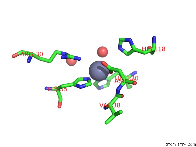
Zinc binding site 1 out of 2 in 5b0h
Go back to
Zinc binding site 1 out
of 2 in the Crystal Structure of Human Leukocyte Cell-Derived Chemotaxin 2

Mono view
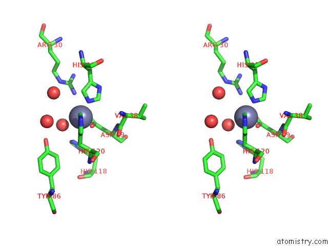
Stereo pair view
Mono view
Stereo pair view
A full contact list of Zinc with other atoms in the Zn binding
site number 1 of Crystal Structure of Human Leukocyte Cell-Derived Chemotaxin 2 within 5.0Å range:
|
Zinc binding site 2 out of 2 in 5b0h
Go back to
Zinc binding site 2 out
of 2 in the Crystal Structure of Human Leukocyte Cell-Derived Chemotaxin 2
Mono view

Stereo pair view
Mono view
Stereo pair view
A full contact list of Zinc with other atoms in the Zn binding
site number 2 of Crystal Structure of Human Leukocyte Cell-Derived Chemotaxin 2 within 5.0Å range:
|
Reference:
H.Zheng,
T.Miyakawa,
Y.Sawano,
A.Asano,
A.Okumura,
S.Yamagoe,
M.Tanokura.
Crystal Structure of Human Leukocyte Cell-Derived Chemotaxin 2 (LECT2) Reveals A Mechanistic Basis of Functional Evolution in A Mammalian Protein with An M23 Metalloendopeptidase Fold J.Biol.Chem. V. 291 17133 2016.
ISSN: ESSN 1083-351X
PubMed: 27334921
DOI: 10.1074/JBC.M116.720375
Page generated: Sun Oct 27 13:16:44 2024
ISSN: ESSN 1083-351X
PubMed: 27334921
DOI: 10.1074/JBC.M116.720375
Last articles
Zn in 9JYWZn in 9IR4
Zn in 9IR3
Zn in 9GMX
Zn in 9GMW
Zn in 9JEJ
Zn in 9ERF
Zn in 9ERE
Zn in 9EGV
Zn in 9EGW