Zinc in PDB 5am8: Crystal Structure of the Angiotensin-1 Converting Enzyme N- Domain in Complex with Amyloid-Beta 4-10
Enzymatic activity of Crystal Structure of the Angiotensin-1 Converting Enzyme N- Domain in Complex with Amyloid-Beta 4-10
All present enzymatic activity of Crystal Structure of the Angiotensin-1 Converting Enzyme N- Domain in Complex with Amyloid-Beta 4-10:
3.4.15.1;
Protein crystallography data
The structure of Crystal Structure of the Angiotensin-1 Converting Enzyme N- Domain in Complex with Amyloid-Beta 4-10, PDB code: 5am8
was solved by
G.Masuyer,
K.M.Larmuth,
R.G.Douglas,
E.D.Sturrock,
K.R.Acharya,
with X-Ray Crystallography technique. A brief refinement statistics is given in the table below:
Resolution Low / High (Å)
|
113.78 /
1.90
|
Space group
|
P 1
|
Cell size a, b, c (Å), α, β, γ (°)
|
73.449,
101.760,
114.361,
85.23,
86.07,
81.45
|
R / Rfree (%)
|
18.543 /
22.382
|
Other elements in 5am8:
The structure of Crystal Structure of the Angiotensin-1 Converting Enzyme N- Domain in Complex with Amyloid-Beta 4-10 also contains other interesting chemical elements:
Zinc Binding Sites:
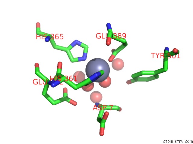
The binding sites of Zinc atom in the Crystal Structure of the Angiotensin-1 Converting Enzyme N- Domain in Complex with Amyloid-Beta 4-10
(pdb code 5am8). This binding sites where shown within
5.0 Angstroms radius around Zinc atom.
In total 4 binding sites of Zinc where determined in the
Crystal Structure of the Angiotensin-1 Converting Enzyme N- Domain in Complex with Amyloid-Beta 4-10, PDB code: 5am8:
Jump to Zinc binding site number:
1;
2;
3;
4;
Zinc binding site 1 out
of 4 in 5am8
Go back to
Zinc Binding Sites List in 5am8
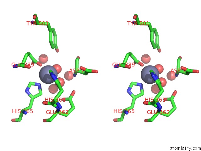
Zinc binding site 1 out
of 4 in the Crystal Structure of the Angiotensin-1 Converting Enzyme N- Domain in Complex with Amyloid-Beta 4-10
Mono view
Stereo pair view
|
A full contact list of Zinc with other atoms in the Zn binding
site number 1 of Crystal Structure of the Angiotensin-1 Converting Enzyme N- Domain in Complex with Amyloid-Beta 4-10 within 5.0Å range:
probe
|
atom
|
residue
|
distance (Å)
|
B
|
Occ
|
A:Zn1001
b:19.4
occ:1.00
|
OE1
|
A:GLU389
|
1.9
|
18.1
|
1.0
|
O
|
A:HOH2289
|
2.0
|
16.3
|
1.0
|
NE2
|
A:HIS361
|
2.0
|
16.9
|
1.0
|
NE2
|
A:HIS365
|
2.1
|
19.6
|
1.0
|
O
|
A:HOH2288
|
2.4
|
29.1
|
1.0
|
CE1
|
A:HIS361
|
2.9
|
17.5
|
1.0
|
CD
|
A:GLU389
|
2.9
|
18.7
|
1.0
|
CE1
|
A:HIS365
|
3.0
|
19.6
|
1.0
|
CD2
|
A:HIS361
|
3.1
|
17.6
|
1.0
|
CD2
|
A:HIS365
|
3.1
|
19.8
|
1.0
|
OE2
|
A:GLU389
|
3.3
|
18.2
|
1.0
|
ND1
|
A:HIS361
|
4.0
|
17.9
|
1.0
|
N
|
P:ASP7
|
4.1
|
25.3
|
1.0
|
ND1
|
A:HIS365
|
4.1
|
19.5
|
1.0
|
CG
|
A:HIS361
|
4.1
|
17.1
|
1.0
|
CE1
|
A:TYR501
|
4.1
|
16.2
|
1.0
|
O
|
A:HOH2266
|
4.2
|
23.2
|
1.0
|
CG
|
A:HIS365
|
4.2
|
19.2
|
1.0
|
CA
|
P:ASP7
|
4.3
|
25.8
|
1.0
|
CG
|
A:GLU389
|
4.3
|
19.3
|
1.0
|
O
|
A:HOH2298
|
4.3
|
18.5
|
1.0
|
OH
|
A:TYR501
|
4.3
|
17.0
|
1.0
|
OE1
|
A:GLU362
|
4.4
|
21.1
|
1.0
|
OE2
|
A:GLU362
|
4.6
|
20.9
|
1.0
|
CA
|
A:GLU389
|
4.6
|
19.9
|
1.0
|
CB
|
A:GLU389
|
4.7
|
19.4
|
1.0
|
CZ
|
A:TYR501
|
4.7
|
16.4
|
1.0
|
C
|
P:ASP7
|
4.8
|
23.1
|
1.0
|
O
|
A:HOH2265
|
4.8
|
28.7
|
1.0
|
CD
|
A:GLU362
|
4.9
|
20.3
|
1.0
|
|
Zinc binding site 2 out
of 4 in 5am8
Go back to
Zinc Binding Sites List in 5am8
Zinc binding site 2 out
of 4 in the Crystal Structure of the Angiotensin-1 Converting Enzyme N- Domain in Complex with Amyloid-Beta 4-10
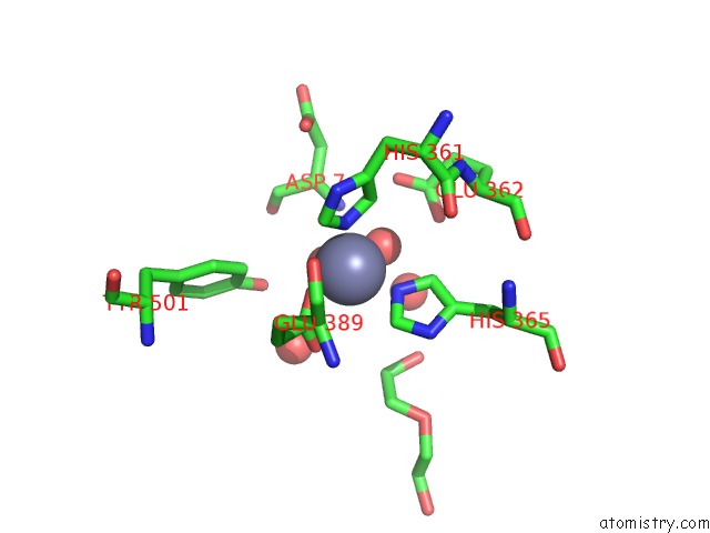
Mono view
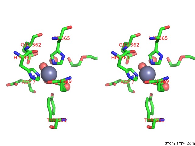
Stereo pair view
|
A full contact list of Zinc with other atoms in the Zn binding
site number 2 of Crystal Structure of the Angiotensin-1 Converting Enzyme N- Domain in Complex with Amyloid-Beta 4-10 within 5.0Å range:
probe
|
atom
|
residue
|
distance (Å)
|
B
|
Occ
|
B:Zn1001
b:16.5
occ:1.00
|
OE1
|
B:GLU389
|
1.9
|
14.1
|
1.0
|
NE2
|
B:HIS361
|
2.1
|
14.5
|
1.0
|
O
|
B:HOH2324
|
2.1
|
15.6
|
1.0
|
NE2
|
B:HIS365
|
2.1
|
16.5
|
1.0
|
O
|
B:HOH2323
|
2.5
|
29.2
|
1.0
|
CD
|
B:GLU389
|
2.9
|
14.8
|
1.0
|
CE1
|
B:HIS365
|
2.9
|
16.0
|
1.0
|
CD2
|
B:HIS361
|
3.0
|
14.5
|
1.0
|
CE1
|
B:HIS361
|
3.0
|
14.2
|
1.0
|
CD2
|
B:HIS365
|
3.2
|
16.1
|
1.0
|
OE2
|
B:GLU389
|
3.2
|
14.7
|
1.0
|
N
|
Q:ASP7
|
3.8
|
24.8
|
1.0
|
O
|
B:HOH2300
|
4.0
|
27.1
|
1.0
|
ND1
|
B:HIS365
|
4.1
|
16.6
|
1.0
|
ND1
|
B:HIS361
|
4.1
|
14.7
|
1.0
|
CE1
|
B:TYR501
|
4.1
|
14.8
|
1.0
|
CG
|
B:HIS361
|
4.1
|
14.3
|
1.0
|
CA
|
Q:ASP7
|
4.2
|
24.4
|
1.0
|
CG
|
B:HIS365
|
4.2
|
15.8
|
1.0
|
OH
|
B:TYR501
|
4.3
|
15.0
|
1.0
|
CG
|
B:GLU389
|
4.3
|
15.0
|
1.0
|
O
|
B:HOH2334
|
4.5
|
19.4
|
1.0
|
OE1
|
B:GLU362
|
4.5
|
17.6
|
1.0
|
OE2
|
B:GLU362
|
4.5
|
17.2
|
1.0
|
CA
|
B:GLU389
|
4.6
|
15.5
|
1.0
|
CZ
|
B:TYR501
|
4.7
|
14.9
|
1.0
|
O
|
B:HOH2299
|
4.7
|
34.7
|
1.0
|
CB
|
B:GLU389
|
4.7
|
15.3
|
1.0
|
C
|
Q:ASP7
|
4.7
|
20.8
|
1.0
|
CD
|
B:GLU362
|
4.8
|
16.8
|
1.0
|
C4
|
B:PEG1205
|
4.9
|
51.8
|
1.0
|
|
Zinc binding site 3 out
of 4 in 5am8
Go back to
Zinc Binding Sites List in 5am8
Zinc binding site 3 out
of 4 in the Crystal Structure of the Angiotensin-1 Converting Enzyme N- Domain in Complex with Amyloid-Beta 4-10
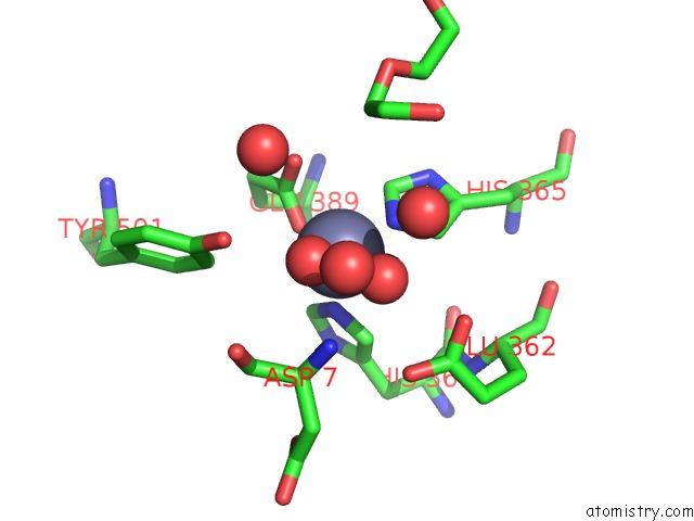
Mono view
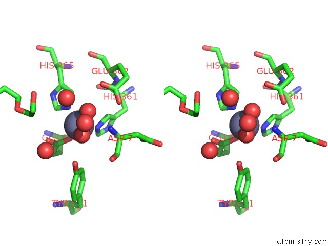
Stereo pair view
|
A full contact list of Zinc with other atoms in the Zn binding
site number 3 of Crystal Structure of the Angiotensin-1 Converting Enzyme N- Domain in Complex with Amyloid-Beta 4-10 within 5.0Å range:
probe
|
atom
|
residue
|
distance (Å)
|
B
|
Occ
|
C:Zn1001
b:15.8
occ:1.00
|
OE1
|
C:GLU389
|
1.9
|
14.0
|
1.0
|
NE2
|
C:HIS365
|
2.0
|
12.7
|
1.0
|
O
|
C:HOH2362
|
2.0
|
10.9
|
1.0
|
NE2
|
C:HIS361
|
2.1
|
14.2
|
1.0
|
O
|
C:HOH2361
|
2.4
|
29.4
|
1.0
|
CD
|
C:GLU389
|
2.9
|
14.1
|
1.0
|
CE1
|
C:HIS365
|
2.9
|
13.2
|
1.0
|
CD2
|
C:HIS361
|
3.0
|
14.0
|
1.0
|
CE1
|
C:HIS361
|
3.1
|
14.4
|
1.0
|
CD2
|
C:HIS365
|
3.1
|
13.1
|
1.0
|
OE2
|
C:GLU389
|
3.2
|
14.1
|
1.0
|
N
|
R:ASP7
|
3.9
|
26.5
|
1.0
|
ND1
|
C:HIS365
|
4.1
|
13.2
|
1.0
|
CE1
|
C:TYR501
|
4.1
|
13.1
|
1.0
|
ND1
|
C:HIS361
|
4.1
|
13.8
|
1.0
|
CG
|
C:HIS361
|
4.1
|
13.6
|
1.0
|
OH
|
C:TYR501
|
4.2
|
12.9
|
1.0
|
CG
|
C:HIS365
|
4.2
|
13.3
|
1.0
|
CG
|
C:GLU389
|
4.2
|
14.1
|
1.0
|
O
|
C:HOH2334
|
4.3
|
30.4
|
1.0
|
CA
|
R:ASP7
|
4.3
|
24.8
|
1.0
|
CA
|
C:GLU389
|
4.5
|
13.9
|
1.0
|
O
|
C:HOH2372
|
4.5
|
14.1
|
1.0
|
OE2
|
C:GLU362
|
4.6
|
15.0
|
1.0
|
CB
|
C:GLU389
|
4.6
|
14.1
|
1.0
|
OE1
|
C:GLU362
|
4.6
|
15.9
|
1.0
|
CZ
|
C:TYR501
|
4.6
|
13.2
|
1.0
|
C
|
R:ASP7
|
4.8
|
21.5
|
1.0
|
C3
|
C:PEG1203
|
4.9
|
44.4
|
1.0
|
CD
|
C:GLU362
|
4.9
|
15.2
|
1.0
|
O
|
C:HOH2333
|
4.9
|
33.3
|
1.0
|
|
Zinc binding site 4 out
of 4 in 5am8
Go back to
Zinc Binding Sites List in 5am8
Zinc binding site 4 out
of 4 in the Crystal Structure of the Angiotensin-1 Converting Enzyme N- Domain in Complex with Amyloid-Beta 4-10
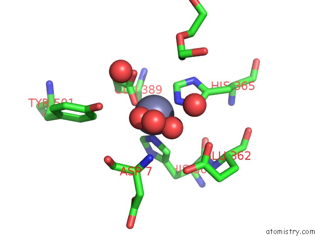
Mono view
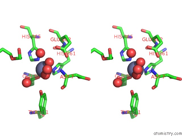
Stereo pair view
|
A full contact list of Zinc with other atoms in the Zn binding
site number 4 of Crystal Structure of the Angiotensin-1 Converting Enzyme N- Domain in Complex with Amyloid-Beta 4-10 within 5.0Å range:
probe
|
atom
|
residue
|
distance (Å)
|
B
|
Occ
|
D:Zn1001
b:15.3
occ:1.00
|
OE1
|
D:GLU389
|
1.9
|
16.2
|
1.0
|
NE2
|
D:HIS361
|
2.0
|
14.8
|
1.0
|
NE2
|
D:HIS365
|
2.0
|
15.9
|
1.0
|
O
|
D:HOH2335
|
2.0
|
11.4
|
1.0
|
O
|
D:HOH2334
|
2.2
|
34.5
|
1.0
|
CD
|
D:GLU389
|
2.9
|
15.3
|
1.0
|
CE1
|
D:HIS365
|
3.0
|
15.5
|
1.0
|
CE1
|
D:HIS361
|
3.0
|
14.2
|
1.0
|
CD2
|
D:HIS361
|
3.0
|
14.2
|
1.0
|
CD2
|
D:HIS365
|
3.1
|
16.0
|
1.0
|
OE2
|
D:GLU389
|
3.2
|
15.3
|
1.0
|
N
|
S:ASP7
|
3.9
|
24.2
|
1.0
|
O
|
D:HOH2313
|
4.0
|
28.8
|
1.0
|
ND1
|
D:HIS361
|
4.1
|
14.4
|
1.0
|
ND1
|
D:HIS365
|
4.1
|
16.1
|
1.0
|
CG
|
D:HIS361
|
4.1
|
14.1
|
1.0
|
CE1
|
D:TYR501
|
4.2
|
12.9
|
1.0
|
CG
|
D:HIS365
|
4.2
|
15.5
|
1.0
|
CG
|
D:GLU389
|
4.3
|
15.4
|
1.0
|
CA
|
S:ASP7
|
4.3
|
24.4
|
1.0
|
OH
|
D:TYR501
|
4.3
|
13.6
|
1.0
|
O
|
D:HOH2346
|
4.4
|
15.7
|
1.0
|
OE1
|
D:GLU362
|
4.5
|
16.7
|
1.0
|
CA
|
D:GLU389
|
4.6
|
15.0
|
1.0
|
OE2
|
D:GLU362
|
4.6
|
15.5
|
1.0
|
O
|
D:HOH2312
|
4.7
|
39.0
|
1.0
|
CB
|
D:GLU389
|
4.7
|
14.8
|
1.0
|
CZ
|
D:TYR501
|
4.7
|
12.9
|
1.0
|
C
|
S:ASP7
|
4.8
|
21.6
|
1.0
|
C3
|
D:PEG1204
|
4.8
|
49.5
|
1.0
|
CD
|
D:GLU362
|
4.9
|
16.0
|
1.0
|
|
Reference:
K.M.Larmuth,
G.Masuyer,
R.G.Douglas,
E.D.Sturrock,
K.R.Acharya.
The Kinetic and Structural Characterisation of Amyloid-Beta Metabolism By Human Angiotensin-1- Converting Enzyme (Ace) Febs J. V. 283 1060 2016.
ISSN: ISSN 1742-464X
PubMed: 26748546
DOI: 10.1111/FEBS.13647
Page generated: Sun Oct 27 13:04:58 2024
|