Zinc »
PDB 4zga-4zvl »
4zr1 »
Zinc in PDB 4zr1: Hydroxylase Domain of SCS7P
Protein crystallography data
The structure of Hydroxylase Domain of SCS7P, PDB code: 4zr1
was solved by
G.Zhu,
M.Koszelak-Rosenblum,
M.G.Malkowski,
Membrane Protein Structuralbiology Consortium (Mpsbc),
with X-Ray Crystallography technique. A brief refinement statistics is given in the table below:
| Resolution Low / High (Å) | 47.79 / 2.60 |
| Space group | I 21 3 |
| Cell size a, b, c (Å), α, β, γ (°) | 202.762, 202.762, 202.762, 90.00, 90.00, 90.00 |
| R / Rfree (%) | 19.7 / 23.9 |
Zinc Binding Sites:
The binding sites of Zinc atom in the Hydroxylase Domain of SCS7P
(pdb code 4zr1). This binding sites where shown within
5.0 Angstroms radius around Zinc atom.
In total 4 binding sites of Zinc where determined in the Hydroxylase Domain of SCS7P, PDB code: 4zr1:
Jump to Zinc binding site number: 1; 2; 3; 4;
In total 4 binding sites of Zinc where determined in the Hydroxylase Domain of SCS7P, PDB code: 4zr1:
Jump to Zinc binding site number: 1; 2; 3; 4;
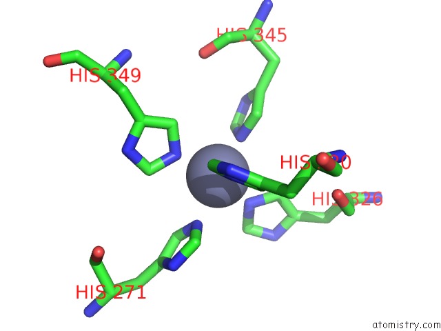
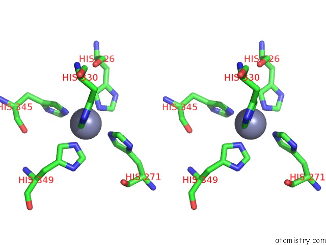
Zinc binding site 1 out of 4 in 4zr1
Go back to
Zinc binding site 1 out
of 4 in the Hydroxylase Domain of SCS7P
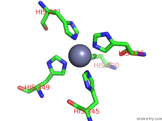
Mono view
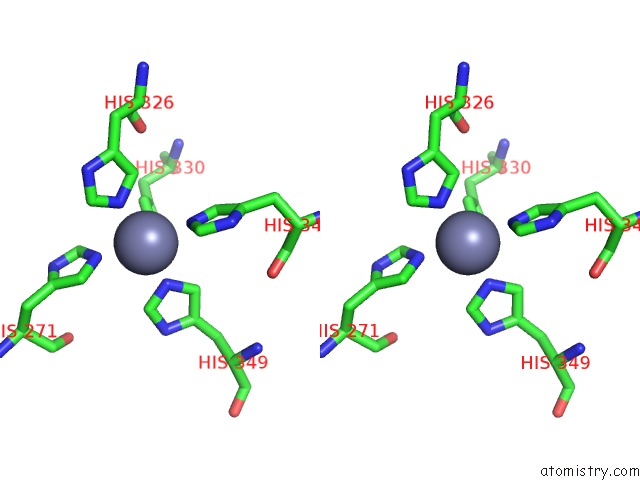
Stereo pair view
Mono view
Stereo pair view
A full contact list of Zinc with other atoms in the Zn binding
site number 1 of Hydroxylase Domain of SCS7P within 5.0Å range:
|
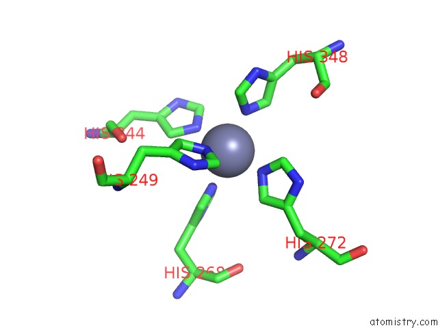
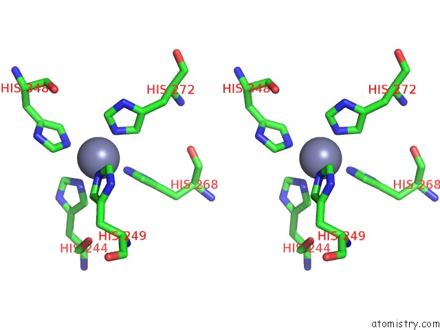
Zinc binding site 2 out of 4 in 4zr1
Go back to
Zinc binding site 2 out
of 4 in the Hydroxylase Domain of SCS7P
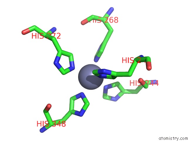
Mono view
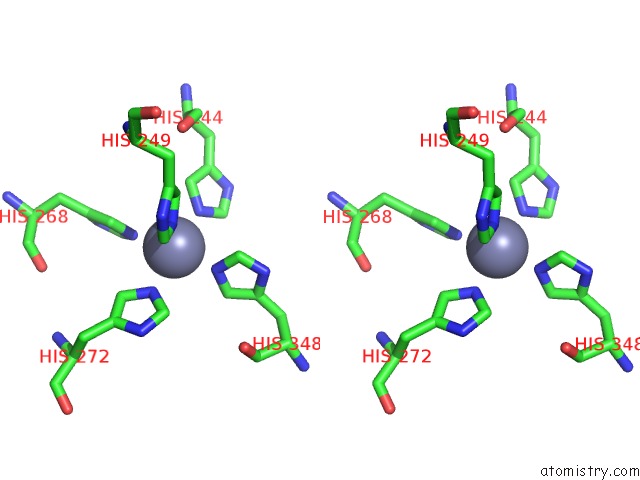
Stereo pair view
Mono view
Stereo pair view
A full contact list of Zinc with other atoms in the Zn binding
site number 2 of Hydroxylase Domain of SCS7P within 5.0Å range:
|
Zinc binding site 3 out of 4 in 4zr1
Go back to
Zinc binding site 3 out
of 4 in the Hydroxylase Domain of SCS7P
Mono view
Stereo pair view
Mono view
Stereo pair view
A full contact list of Zinc with other atoms in the Zn binding
site number 3 of Hydroxylase Domain of SCS7P within 5.0Å range:
|
Zinc binding site 4 out of 4 in 4zr1
Go back to
Zinc binding site 4 out
of 4 in the Hydroxylase Domain of SCS7P
Mono view
Stereo pair view
Mono view
Stereo pair view
A full contact list of Zinc with other atoms in the Zn binding
site number 4 of Hydroxylase Domain of SCS7P within 5.0Å range:
|
Reference:
G.Zhu,
M.Koszelak-Rosenblum,
S.M.Connelly,
M.E.Dumont,
M.G.Malkowski.
The Crystal Structure of An Integral Membrane Fatty Acid Alpha-Hydroxylase. J.Biol.Chem. V. 290 29820 2015.
ISSN: ESSN 1083-351X
PubMed: 26515067
DOI: 10.1074/JBC.M115.680124
Page generated: Sun Oct 27 11:55:35 2024
ISSN: ESSN 1083-351X
PubMed: 26515067
DOI: 10.1074/JBC.M115.680124
Last articles
Zn in 9JYWZn in 9IR4
Zn in 9IR3
Zn in 9GMX
Zn in 9GMW
Zn in 9JEJ
Zn in 9ERF
Zn in 9ERE
Zn in 9EGV
Zn in 9EGW