Zinc »
PDB 4ylp-4z0q »
4ymk »
Zinc in PDB 4ymk: Crystal Structure of Stearoyl-Coenzyme A Desaturase 1
Enzymatic activity of Crystal Structure of Stearoyl-Coenzyme A Desaturase 1
All present enzymatic activity of Crystal Structure of Stearoyl-Coenzyme A Desaturase 1:
1.14.19.1;
1.14.19.1;
Protein crystallography data
The structure of Crystal Structure of Stearoyl-Coenzyme A Desaturase 1, PDB code: 4ymk
was solved by
Y.Bai,
J.G.Mccoy,
K.R.Rajashankar,
M.Zhou,
with X-Ray Crystallography technique. A brief refinement statistics is given in the table below:
| Resolution Low / High (Å) | 47.41 / 2.61 |
| Space group | P 21 21 21 |
| Cell size a, b, c (Å), α, β, γ (°) | 77.061, 113.766, 141.698, 90.00, 90.00, 90.00 |
| R / Rfree (%) | 20.3 / 23.5 |
Zinc Binding Sites:
The binding sites of Zinc atom in the Crystal Structure of Stearoyl-Coenzyme A Desaturase 1
(pdb code 4ymk). This binding sites where shown within
5.0 Angstroms radius around Zinc atom.
In total 4 binding sites of Zinc where determined in the Crystal Structure of Stearoyl-Coenzyme A Desaturase 1, PDB code: 4ymk:
Jump to Zinc binding site number: 1; 2; 3; 4;
In total 4 binding sites of Zinc where determined in the Crystal Structure of Stearoyl-Coenzyme A Desaturase 1, PDB code: 4ymk:
Jump to Zinc binding site number: 1; 2; 3; 4;
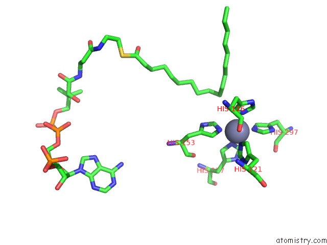
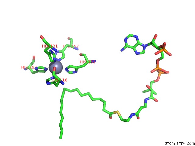
Zinc binding site 1 out of 4 in 4ymk
Go back to
Zinc binding site 1 out
of 4 in the Crystal Structure of Stearoyl-Coenzyme A Desaturase 1
Mono view
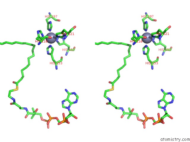
Stereo pair view
Mono view
Stereo pair view
A full contact list of Zinc with other atoms in the Zn binding
site number 1 of Crystal Structure of Stearoyl-Coenzyme A Desaturase 1 within 5.0Å range:
|
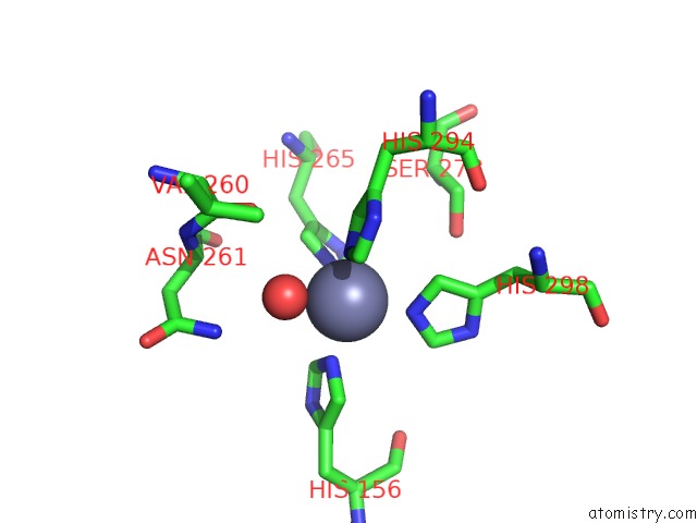
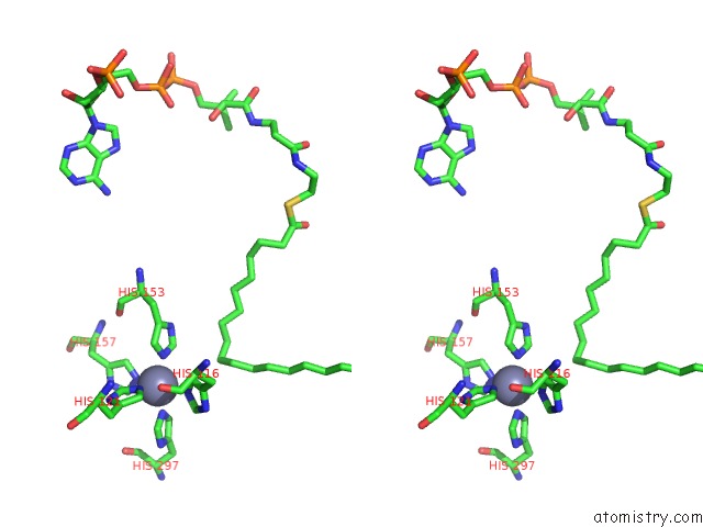
Zinc binding site 2 out of 4 in 4ymk
Go back to
Zinc binding site 2 out
of 4 in the Crystal Structure of Stearoyl-Coenzyme A Desaturase 1
Mono view
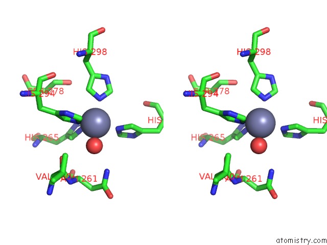
Stereo pair view
Mono view
Stereo pair view
A full contact list of Zinc with other atoms in the Zn binding
site number 2 of Crystal Structure of Stearoyl-Coenzyme A Desaturase 1 within 5.0Å range:
|
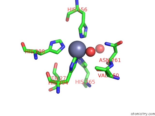
Zinc binding site 3 out of 4 in 4ymk
Go back to
Zinc binding site 3 out
of 4 in the Crystal Structure of Stearoyl-Coenzyme A Desaturase 1
Mono view
Stereo pair view
Mono view
Stereo pair view
A full contact list of Zinc with other atoms in the Zn binding
site number 3 of Crystal Structure of Stearoyl-Coenzyme A Desaturase 1 within 5.0Å range:
|
Zinc binding site 4 out of 4 in 4ymk
Go back to
Zinc binding site 4 out
of 4 in the Crystal Structure of Stearoyl-Coenzyme A Desaturase 1
Mono view
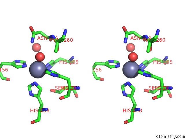
Stereo pair view
Mono view
Stereo pair view
A full contact list of Zinc with other atoms in the Zn binding
site number 4 of Crystal Structure of Stearoyl-Coenzyme A Desaturase 1 within 5.0Å range:
|
Reference:
Y.Bai,
J.G.Mccoy,
E.J.Levin,
P.Sobrado,
K.R.Rajashankar,
B.G.Fox,
M.Zhou.
X-Ray Structure of A Mammalian Stearoyl-Coa Desaturase. Nature V. 524 252 2015.
ISSN: ESSN 1476-4687
PubMed: 26098370
DOI: 10.1038/NATURE14549
Page generated: Sun Oct 27 11:19:51 2024
ISSN: ESSN 1476-4687
PubMed: 26098370
DOI: 10.1038/NATURE14549
Last articles
Zn in 9JYWZn in 9IR4
Zn in 9IR3
Zn in 9GMX
Zn in 9GMW
Zn in 9JEJ
Zn in 9ERF
Zn in 9ERE
Zn in 9EGV
Zn in 9EGW