Zinc »
PDB 4qqr-4r5t »
4r5t »
Zinc in PDB 4r5t: Structure of the M1 Alanylaminopeptidase From Malaria Complexed with A Hydroxamic Acid-Based Inhibitor
Protein crystallography data
The structure of Structure of the M1 Alanylaminopeptidase From Malaria Complexed with A Hydroxamic Acid-Based Inhibitor, PDB code: 4r5t
was solved by
N.Drinkwater,
S.Mcgowan,
with X-Ray Crystallography technique. A brief refinement statistics is given in the table below:
| Resolution Low / High (Å) | 35.84 / 1.98 |
| Space group | P 21 21 21 |
| Cell size a, b, c (Å), α, β, γ (°) | 75.193, 109.587, 118.490, 90.00, 90.00, 90.00 |
| R / Rfree (%) | 18.3 / 22.8 |
Zinc Binding Sites:
The binding sites of Zinc atom in the Structure of the M1 Alanylaminopeptidase From Malaria Complexed with A Hydroxamic Acid-Based Inhibitor
(pdb code 4r5t). This binding sites where shown within
5.0 Angstroms radius around Zinc atom.
In total only one binding site of Zinc was determined in the Structure of the M1 Alanylaminopeptidase From Malaria Complexed with A Hydroxamic Acid-Based Inhibitor, PDB code: 4r5t:
In total only one binding site of Zinc was determined in the Structure of the M1 Alanylaminopeptidase From Malaria Complexed with A Hydroxamic Acid-Based Inhibitor, PDB code: 4r5t:
Zinc binding site 1 out of 1 in 4r5t
Go back to
Zinc binding site 1 out
of 1 in the Structure of the M1 Alanylaminopeptidase From Malaria Complexed with A Hydroxamic Acid-Based Inhibitor
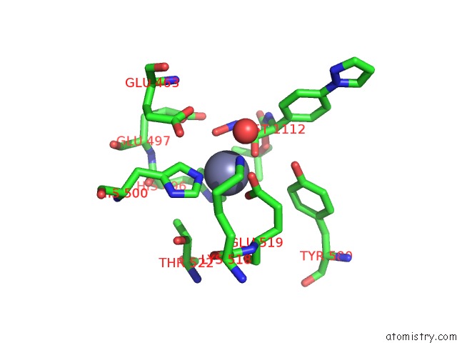
Mono view
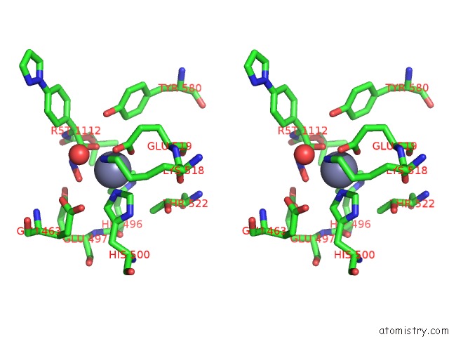
Stereo pair view
Mono view
Stereo pair view
A full contact list of Zinc with other atoms in the Zn binding
site number 1 of Structure of the M1 Alanylaminopeptidase From Malaria Complexed with A Hydroxamic Acid-Based Inhibitor within 5.0Å range:
|
Reference:
S.N.Mistry,
N.Drinkwater,
C.Ruggeri,
K.Kannan Sivaraman,
S.Loganathan,
S.Fletcher,
M.Drag,
A.Paiardini,
V.M.Avery,
P.J.Scammells,
S.Mcgowan.
A Two-Pronged Attack: Dual Inhibition of Plasmodium Falciparum M1 and M17 Metalloaminopeptidases By A Novel Series of Hydroxamic Acid-Based Inhibitors. J.Med.Chem. 2014.
ISSN: ISSN 0022-2623
PubMed: 25299353
DOI: 10.1021/JM501323A
Page generated: Sun Oct 27 06:52:55 2024
ISSN: ISSN 0022-2623
PubMed: 25299353
DOI: 10.1021/JM501323A
Last articles
Zn in 9JYWZn in 9IR4
Zn in 9IR3
Zn in 9GMX
Zn in 9GMW
Zn in 9JEJ
Zn in 9ERF
Zn in 9ERE
Zn in 9EGV
Zn in 9EGW