Zinc »
PDB 4qqr-4r5t »
4quo »
Zinc in PDB 4quo: Crystal Structure of Aminopeptidase N in Complex with the Phosphinic Dipeptide Analogue Ll-(R,S)-Hphep[CH2]Phe(3-CH2NH2)
Enzymatic activity of Crystal Structure of Aminopeptidase N in Complex with the Phosphinic Dipeptide Analogue Ll-(R,S)-Hphep[CH2]Phe(3-CH2NH2)
All present enzymatic activity of Crystal Structure of Aminopeptidase N in Complex with the Phosphinic Dipeptide Analogue Ll-(R,S)-Hphep[CH2]Phe(3-CH2NH2):
3.4.11.2;
3.4.11.2;
Protein crystallography data
The structure of Crystal Structure of Aminopeptidase N in Complex with the Phosphinic Dipeptide Analogue Ll-(R,S)-Hphep[CH2]Phe(3-CH2NH2), PDB code: 4quo
was solved by
B.Nocek,
R.Mulligan,
A.Joachimak,
S.Vassiliou,
L.Berlicki,
A.Mucha,
with X-Ray Crystallography technique. A brief refinement statistics is given in the table below:
| Resolution Low / High (Å) | 27.98 / 1.65 |
| Space group | H 3 |
| Cell size a, b, c (Å), α, β, γ (°) | 223.707, 223.707, 57.769, 90.00, 90.00, 120.00 |
| R / Rfree (%) | 14.8 / 18.2 |
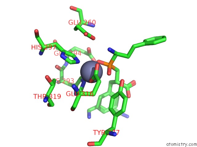
Zinc Binding Sites:
The binding sites of Zinc atom in the Crystal Structure of Aminopeptidase N in Complex with the Phosphinic Dipeptide Analogue Ll-(R,S)-Hphep[CH2]Phe(3-CH2NH2)
(pdb code 4quo). This binding sites where shown within
5.0 Angstroms radius around Zinc atom.
In total only one binding site of Zinc was determined in the Crystal Structure of Aminopeptidase N in Complex with the Phosphinic Dipeptide Analogue Ll-(R,S)-Hphep[CH2]Phe(3-CH2NH2), PDB code: 4quo:
In total only one binding site of Zinc was determined in the Crystal Structure of Aminopeptidase N in Complex with the Phosphinic Dipeptide Analogue Ll-(R,S)-Hphep[CH2]Phe(3-CH2NH2), PDB code: 4quo:
Zinc binding site 1 out of 1 in 4quo
Go back to
Zinc binding site 1 out
of 1 in the Crystal Structure of Aminopeptidase N in Complex with the Phosphinic Dipeptide Analogue Ll-(R,S)-Hphep[CH2]Phe(3-CH2NH2)
Mono view

Stereo pair view
Mono view
Stereo pair view
A full contact list of Zinc with other atoms in the Zn binding
site number 1 of Crystal Structure of Aminopeptidase N in Complex with the Phosphinic Dipeptide Analogue Ll-(R,S)-Hphep[CH2]Phe(3-CH2NH2) within 5.0Å range:
|
Reference:
S.Vassiliou,
E.Weglarz-Tomczak,
L.Berlicki,
M.Paweczak,
B.Nocek,
R.Mulligan,
A.Joachimiak,
A.Mucha.
Structure-Guided, Single-Point Modifications in the Phosphinic Dipeptide Structure Yield Highly Potent and Selective Inhibitors of Neutral Aminopeptidases. J.Med.Chem. V. 57 8140 2014.
ISSN: ISSN 0022-2623
PubMed: 25192493
DOI: 10.1021/JM501071F
Page generated: Sun Oct 27 06:45:20 2024
ISSN: ISSN 0022-2623
PubMed: 25192493
DOI: 10.1021/JM501071F
Last articles
Zn in 9JYWZn in 9IR4
Zn in 9IR3
Zn in 9GMX
Zn in 9GMW
Zn in 9JEJ
Zn in 9ERF
Zn in 9ERE
Zn in 9EGV
Zn in 9EGW