Zinc »
PDB 4lep-4llg »
4lk1 »
Zinc in PDB 4lk1: Crystal Structure Analysis of the E.Coli Holoenzyme
Enzymatic activity of Crystal Structure Analysis of the E.Coli Holoenzyme
All present enzymatic activity of Crystal Structure Analysis of the E.Coli Holoenzyme:
2.7.7.6;
2.7.7.6;
Protein crystallography data
The structure of Crystal Structure Analysis of the E.Coli Holoenzyme, PDB code: 4lk1
was solved by
B.Bae,
S.A.Darst,
with X-Ray Crystallography technique. A brief refinement statistics is given in the table below:
| Resolution Low / High (Å) | 44.78 / 3.84 |
| Space group | P 21 21 21 |
| Cell size a, b, c (Å), α, β, γ (°) | 186.675, 206.392, 308.836, 90.00, 90.00, 90.00 |
| R / Rfree (%) | 22.4 / 27.2 |
Other elements in 4lk1:
The structure of Crystal Structure Analysis of the E.Coli Holoenzyme also contains other interesting chemical elements:
| Magnesium | (Mg) | 2 atoms |
Zinc Binding Sites:
The binding sites of Zinc atom in the Crystal Structure Analysis of the E.Coli Holoenzyme
(pdb code 4lk1). This binding sites where shown within
5.0 Angstroms radius around Zinc atom.
In total 4 binding sites of Zinc where determined in the Crystal Structure Analysis of the E.Coli Holoenzyme, PDB code: 4lk1:
Jump to Zinc binding site number: 1; 2; 3; 4;
In total 4 binding sites of Zinc where determined in the Crystal Structure Analysis of the E.Coli Holoenzyme, PDB code: 4lk1:
Jump to Zinc binding site number: 1; 2; 3; 4;
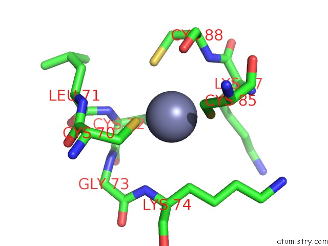
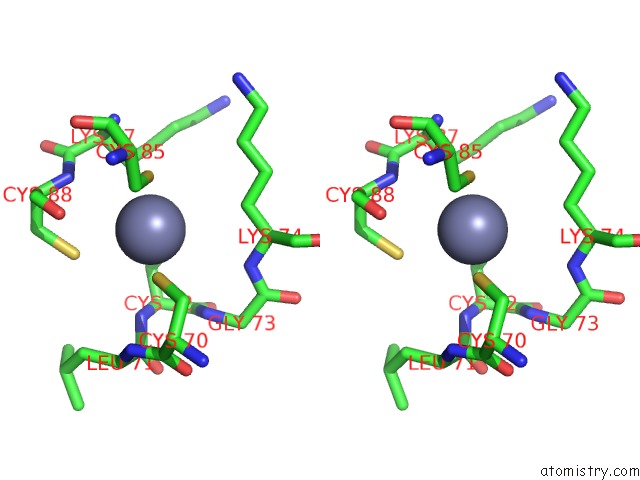
Zinc binding site 1 out of 4 in 4lk1
Go back to
Zinc binding site 1 out
of 4 in the Crystal Structure Analysis of the E.Coli Holoenzyme
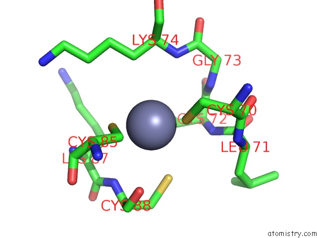
Mono view
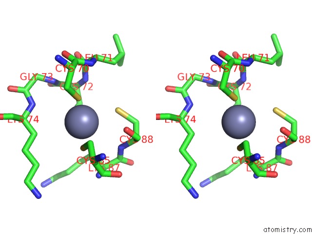
Stereo pair view
Mono view
Stereo pair view
A full contact list of Zinc with other atoms in the Zn binding
site number 1 of Crystal Structure Analysis of the E.Coli Holoenzyme within 5.0Å range:
|
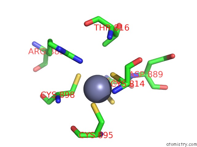
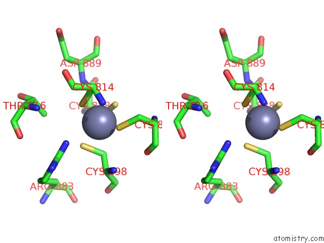
Zinc binding site 2 out of 4 in 4lk1
Go back to
Zinc binding site 2 out
of 4 in the Crystal Structure Analysis of the E.Coli Holoenzyme
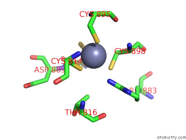
Mono view
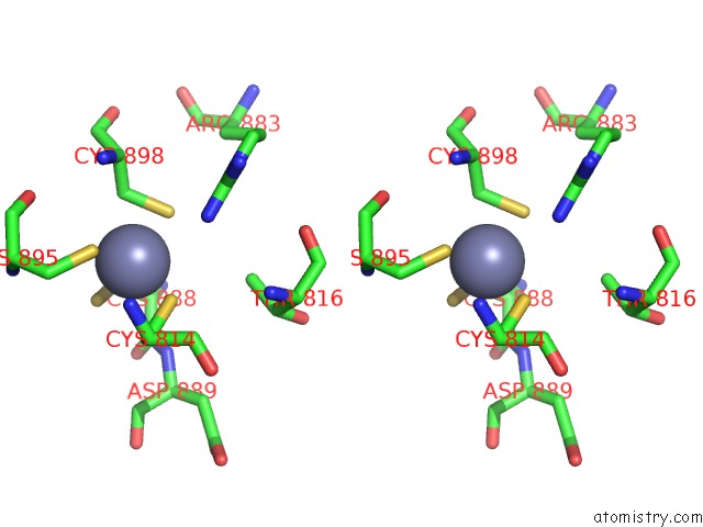
Stereo pair view
Mono view
Stereo pair view
A full contact list of Zinc with other atoms in the Zn binding
site number 2 of Crystal Structure Analysis of the E.Coli Holoenzyme within 5.0Å range:
|
Zinc binding site 3 out of 4 in 4lk1
Go back to
Zinc binding site 3 out
of 4 in the Crystal Structure Analysis of the E.Coli Holoenzyme
Mono view
Stereo pair view
Mono view
Stereo pair view
A full contact list of Zinc with other atoms in the Zn binding
site number 3 of Crystal Structure Analysis of the E.Coli Holoenzyme within 5.0Å range:
|
Zinc binding site 4 out of 4 in 4lk1
Go back to
Zinc binding site 4 out
of 4 in the Crystal Structure Analysis of the E.Coli Holoenzyme
Mono view
Stereo pair view
Mono view
Stereo pair view
A full contact list of Zinc with other atoms in the Zn binding
site number 4 of Crystal Structure Analysis of the E.Coli Holoenzyme within 5.0Å range:
|
Reference:
B.Bae,
E.Davis,
D.Brown,
E.A.Campbell,
S.Wigneshweraraj,
S.A.Darst.
Phage T7 GP2 Inhibition of Escherichia Coli Rna Polymerase Involves Misappropriation of Sigma 70 Domain 1.1. Proc.Natl.Acad.Sci.Usa V. 110 19772 2013.
ISSN: ISSN 0027-8424
PubMed: 24218560
DOI: 10.1073/PNAS.1314576110
Page generated: Sun Oct 27 01:54:35 2024
ISSN: ISSN 0027-8424
PubMed: 24218560
DOI: 10.1073/PNAS.1314576110
Last articles
Zn in 9JYWZn in 9IR4
Zn in 9IR3
Zn in 9GMX
Zn in 9GMW
Zn in 9JEJ
Zn in 9ERF
Zn in 9ERE
Zn in 9EGV
Zn in 9EGW