Zinc »
PDB 4lep-4llg »
4lew »
Zinc in PDB 4lew: Structure of Human Cgas
Protein crystallography data
The structure of Structure of Human Cgas, PDB code: 4lew
was solved by
P.Li,
with X-Ray Crystallography technique. A brief refinement statistics is given in the table below:
| Resolution Low / High (Å) | 40.75 / 2.04 |
| Space group | P 1 21 1 |
| Cell size a, b, c (Å), α, β, γ (°) | 46.815, 111.480, 76.414, 90.00, 92.73, 90.00 |
| R / Rfree (%) | 17.9 / 22.3 |
Zinc Binding Sites:
The binding sites of Zinc atom in the Structure of Human Cgas
(pdb code 4lew). This binding sites where shown within
5.0 Angstroms radius around Zinc atom.
In total 2 binding sites of Zinc where determined in the Structure of Human Cgas, PDB code: 4lew:
Jump to Zinc binding site number: 1; 2;
In total 2 binding sites of Zinc where determined in the Structure of Human Cgas, PDB code: 4lew:
Jump to Zinc binding site number: 1; 2;
Zinc binding site 1 out of 2 in 4lew
Go back to
Zinc binding site 1 out
of 2 in the Structure of Human Cgas
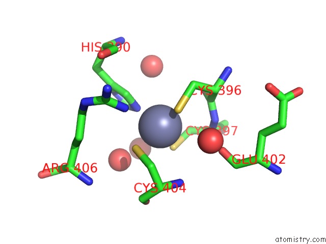
Mono view
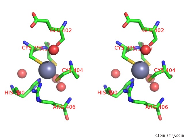
Stereo pair view
Mono view
Stereo pair view
A full contact list of Zinc with other atoms in the Zn binding
site number 1 of Structure of Human Cgas within 5.0Å range:
|
Zinc binding site 2 out of 2 in 4lew
Go back to
Zinc binding site 2 out
of 2 in the Structure of Human Cgas
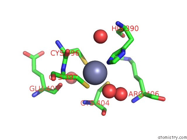
Mono view
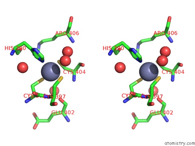
Stereo pair view
Mono view
Stereo pair view
A full contact list of Zinc with other atoms in the Zn binding
site number 2 of Structure of Human Cgas within 5.0Å range:
|
Reference:
X.Li,
C.Shu,
G.Yi,
C.T.Chaton,
C.L.Shelton,
J.Diao,
X.Zuo,
C.C.Kao,
A.B.Herr,
P.Li.
Cyclic Gmp-Amp Synthase Is Activated By Double-Stranded Dna-Induced Oligomerization. Immunity V. 39 1019 2013.
ISSN: ISSN 1074-7613
PubMed: 24332030
DOI: 10.1016/J.IMMUNI.2013.10.019
Page generated: Sun Oct 27 01:47:33 2024
ISSN: ISSN 1074-7613
PubMed: 24332030
DOI: 10.1016/J.IMMUNI.2013.10.019
Last articles
Zn in 9JYWZn in 9IR4
Zn in 9IR3
Zn in 9GMX
Zn in 9GMW
Zn in 9JEJ
Zn in 9ERF
Zn in 9ERE
Zn in 9EGV
Zn in 9EGW