Zinc »
PDB 4k4f-4k98 »
4k95 »
Zinc in PDB 4k95: Crystal Structure of Parkin
Protein crystallography data
The structure of Crystal Structure of Parkin, PDB code: 4k95
was solved by
M.Seirafi,
M.Menade,
V.Sauve,
G.Kozlov,
J.-F.Trempe,
B.Nagar,
K.Gehring,
with X-Ray Crystallography technique. A brief refinement statistics is given in the table below:
| Resolution Low / High (Å) | 49.33 / 6.50 |
| Space group | P 21 21 2 |
| Cell size a, b, c (Å), α, β, γ (°) | 208.598, 277.439, 125.891, 90.00, 90.00, 90.00 |
| R / Rfree (%) | 30.7 / 32.7 |
Zinc Binding Sites:
Pages:
>>> Page 1 <<< Page 2, Binding sites: 11 - 20; Page 3, Binding sites: 21 - 30; Page 4, Binding sites: 31 - 40; Page 5, Binding sites: 41 - 50; Page 6, Binding sites: 51 - 60; Page 7, Binding sites: 61 - 70; Page 8, Binding sites: 71 - 80; Page 9, Binding sites: 81 - 90; Page 10, Binding sites: 91 - 96;Binding sites:
The binding sites of Zinc atom in the Crystal Structure of Parkin (pdb code 4k95). This binding sites where shown within 5.0 Angstroms radius around Zinc atom.In total 96 binding sites of Zinc where determined in the Crystal Structure of Parkin, PDB code: 4k95:
Jump to Zinc binding site number: 1; 2; 3; 4; 5; 6; 7; 8; 9; 10;
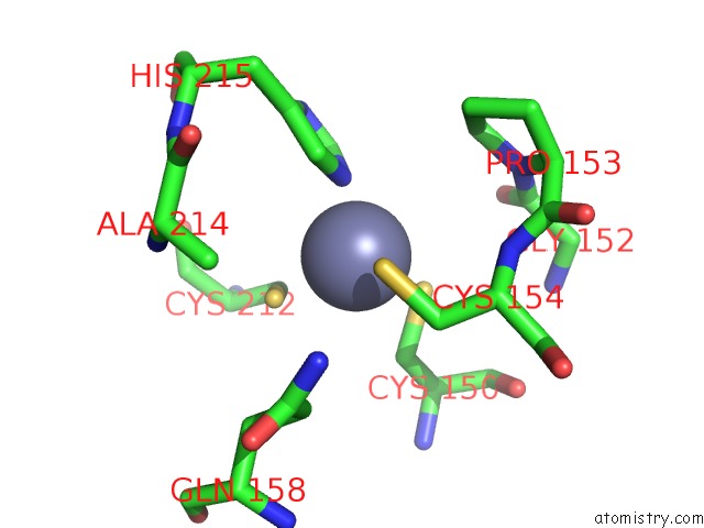
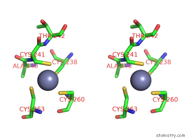
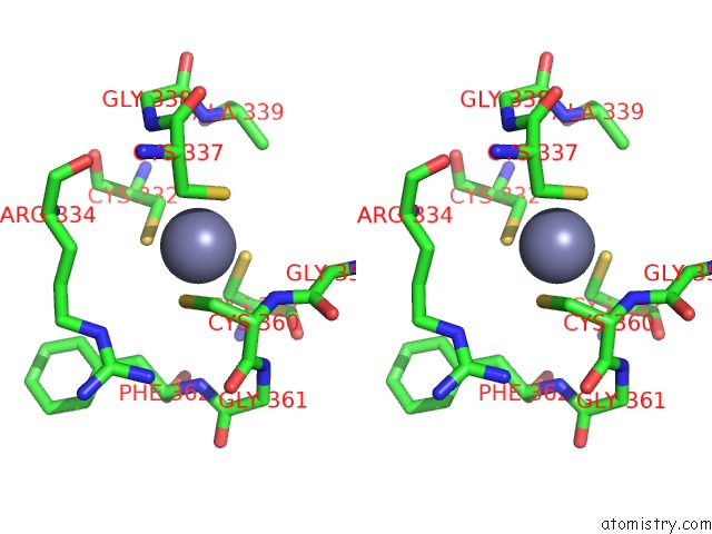
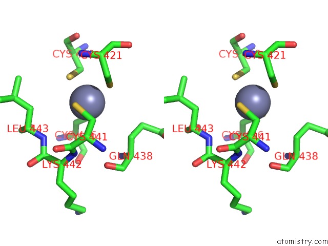
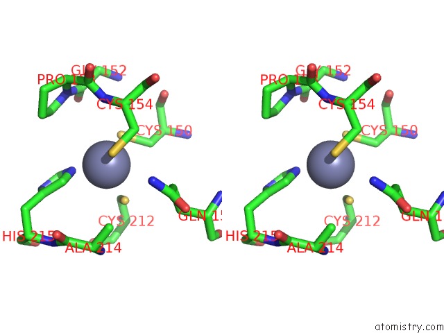
Zinc binding site 1 out of 96 in 4k95
Go back to
Zinc binding site 1 out
of 96 in the Crystal Structure of Parkin
Mono view

Stereo pair view
Mono view
Stereo pair view
A full contact list of Zinc with other atoms in the Zn binding
site number 1 of Crystal Structure of Parkin within 5.0Å range:
|
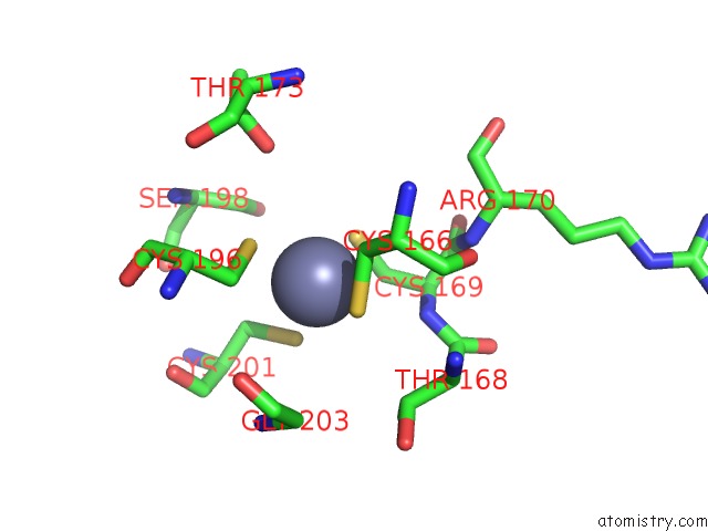
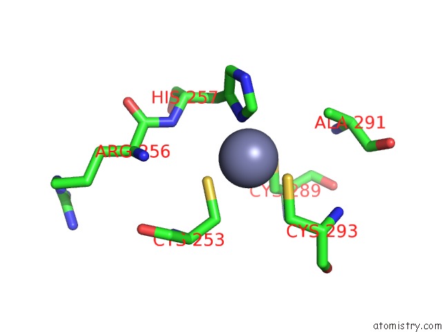
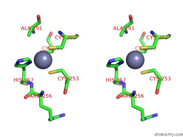
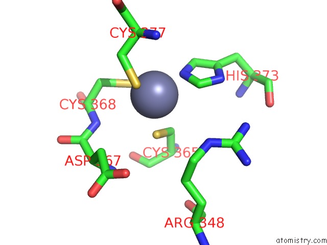
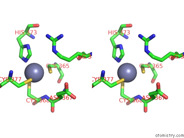
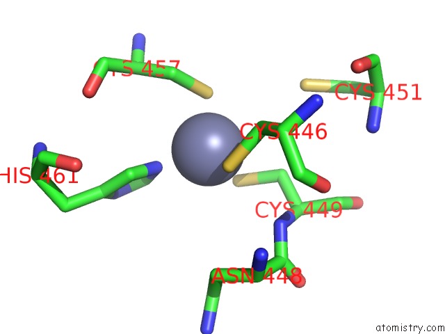
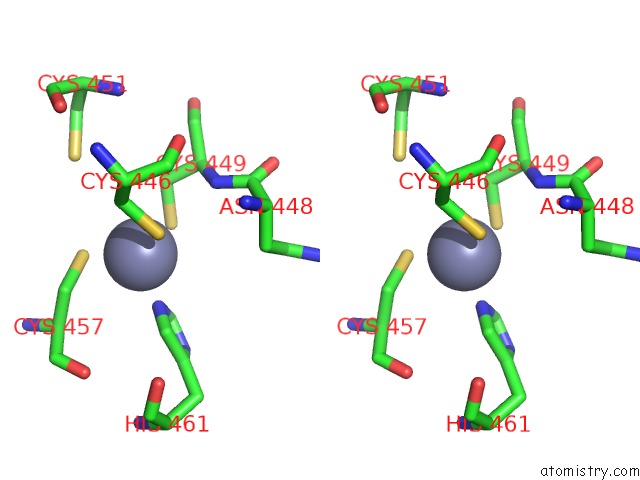
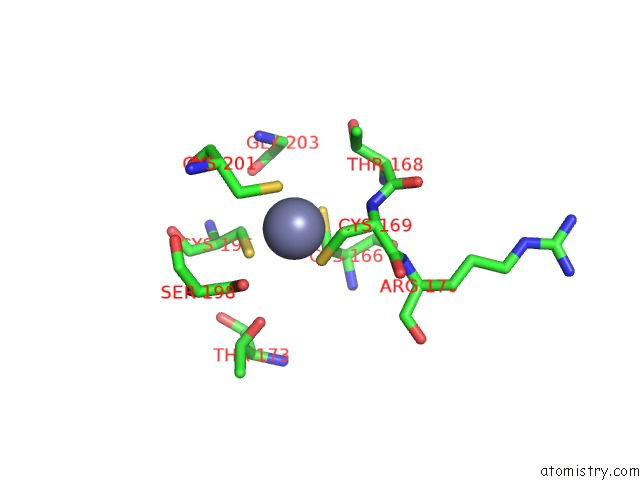
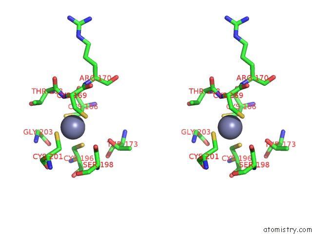
Zinc binding site 2 out of 96 in 4k95
Go back to
Zinc binding site 2 out
of 96 in the Crystal Structure of Parkin
Mono view
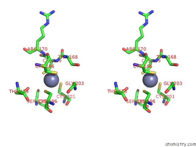
Stereo pair view
Mono view
Stereo pair view
A full contact list of Zinc with other atoms in the Zn binding
site number 2 of Crystal Structure of Parkin within 5.0Å range:
|
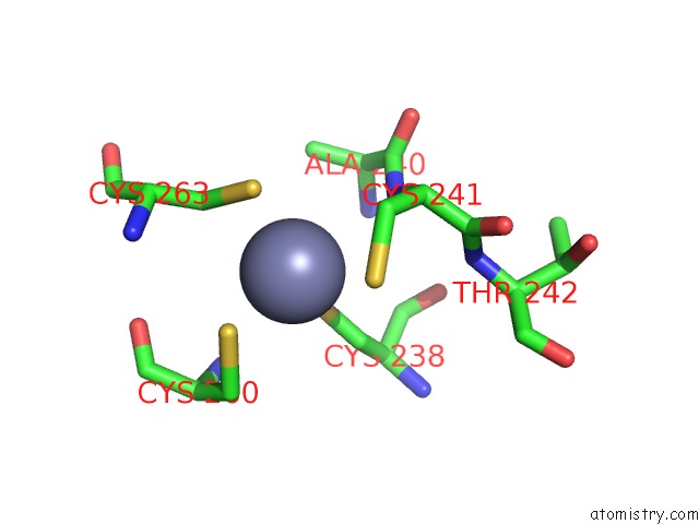
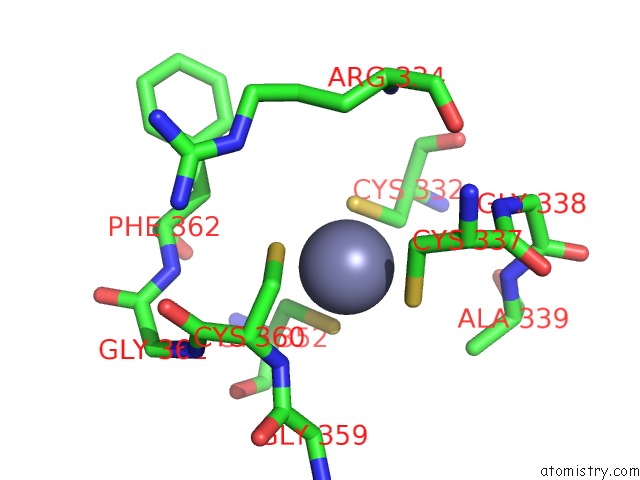
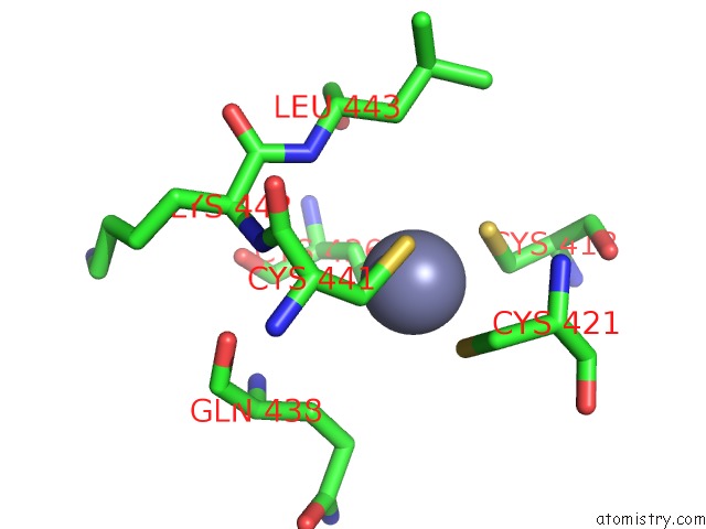
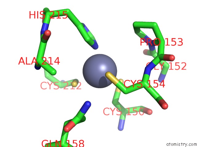
Zinc binding site 3 out of 96 in 4k95
Go back to
Zinc binding site 3 out
of 96 in the Crystal Structure of Parkin
Mono view
Stereo pair view
Mono view
Stereo pair view
A full contact list of Zinc with other atoms in the Zn binding
site number 3 of Crystal Structure of Parkin within 5.0Å range:
|
Zinc binding site 4 out of 96 in 4k95
Go back to
Zinc binding site 4 out
of 96 in the Crystal Structure of Parkin
Mono view
Stereo pair view
Mono view
Stereo pair view
A full contact list of Zinc with other atoms in the Zn binding
site number 4 of Crystal Structure of Parkin within 5.0Å range:
|
Zinc binding site 5 out of 96 in 4k95
Go back to
Zinc binding site 5 out
of 96 in the Crystal Structure of Parkin
Mono view
Stereo pair view
Mono view
Stereo pair view
A full contact list of Zinc with other atoms in the Zn binding
site number 5 of Crystal Structure of Parkin within 5.0Å range:
|
Zinc binding site 6 out of 96 in 4k95
Go back to
Zinc binding site 6 out
of 96 in the Crystal Structure of Parkin
Mono view
Stereo pair view
Mono view
Stereo pair view
A full contact list of Zinc with other atoms in the Zn binding
site number 6 of Crystal Structure of Parkin within 5.0Å range:
|
Zinc binding site 7 out of 96 in 4k95
Go back to
Zinc binding site 7 out
of 96 in the Crystal Structure of Parkin
Mono view
Stereo pair view
Mono view
Stereo pair view
A full contact list of Zinc with other atoms in the Zn binding
site number 7 of Crystal Structure of Parkin within 5.0Å range:
|
Zinc binding site 8 out of 96 in 4k95
Go back to
Zinc binding site 8 out
of 96 in the Crystal Structure of Parkin
Mono view
Stereo pair view
Mono view
Stereo pair view
A full contact list of Zinc with other atoms in the Zn binding
site number 8 of Crystal Structure of Parkin within 5.0Å range:
|
Zinc binding site 9 out of 96 in 4k95
Go back to
Zinc binding site 9 out
of 96 in the Crystal Structure of Parkin
Mono view
Stereo pair view
Mono view
Stereo pair view
A full contact list of Zinc with other atoms in the Zn binding
site number 9 of Crystal Structure of Parkin within 5.0Å range:
|
Zinc binding site 10 out of 96 in 4k95
Go back to
Zinc binding site 10 out
of 96 in the Crystal Structure of Parkin
Mono view
Stereo pair view
Mono view
Stereo pair view
A full contact list of Zinc with other atoms in the Zn binding
site number 10 of Crystal Structure of Parkin within 5.0Å range:
|
Reference:
J.F.Trempe,
V.Sauve,
K.Grenier,
M.Seirafi,
M.Y.Tang,
M.Menade,
S.Al-Abdul-Wahid,
J.Krett,
K.Wong,
G.Kozlov,
B.Nagar,
E.A.Fon,
K.Gehring.
Structure of Parkin Reveals Mechanisms For Ubiquitin Ligase Activation. Science V. 340 1451 2013.
ISSN: ISSN 0036-8075
PubMed: 23661642
DOI: 10.1126/SCIENCE.1237908
Page generated: Sun Oct 27 01:50:35 2024
ISSN: ISSN 0036-8075
PubMed: 23661642
DOI: 10.1126/SCIENCE.1237908
Last articles
Zn in 9JYWZn in 9IR4
Zn in 9IR3
Zn in 9GMX
Zn in 9GMW
Zn in 9JEJ
Zn in 9ERF
Zn in 9ERE
Zn in 9EGV
Zn in 9EGW