Zinc »
PDB 4hk7-4hwp »
4htx »
Zinc in PDB 4htx: Crystal Structure of PDE2 Catalytic Domain in Complex with BAY60-7550
Enzymatic activity of Crystal Structure of PDE2 Catalytic Domain in Complex with BAY60-7550
All present enzymatic activity of Crystal Structure of PDE2 Catalytic Domain in Complex with BAY60-7550:
3.1.4.17;
3.1.4.17;
Protein crystallography data
The structure of Crystal Structure of PDE2 Catalytic Domain in Complex with BAY60-7550, PDB code: 4htx
was solved by
J.Zhu,
Q.Huang,
with X-Ray Crystallography technique. A brief refinement statistics is given in the table below:
| Resolution Low / High (Å) | 44.16 / 1.90 |
| Space group | P 1 |
| Cell size a, b, c (Å), α, β, γ (°) | 56.260, 73.820, 92.470, 109.59, 91.93, 90.88 |
| R / Rfree (%) | 17.3 / 22.7 |
Other elements in 4htx:
The structure of Crystal Structure of PDE2 Catalytic Domain in Complex with BAY60-7550 also contains other interesting chemical elements:
| Magnesium | (Mg) | 4 atoms |
Zinc Binding Sites:
The binding sites of Zinc atom in the Crystal Structure of PDE2 Catalytic Domain in Complex with BAY60-7550
(pdb code 4htx). This binding sites where shown within
5.0 Angstroms radius around Zinc atom.
In total 4 binding sites of Zinc where determined in the Crystal Structure of PDE2 Catalytic Domain in Complex with BAY60-7550, PDB code: 4htx:
Jump to Zinc binding site number: 1; 2; 3; 4;
In total 4 binding sites of Zinc where determined in the Crystal Structure of PDE2 Catalytic Domain in Complex with BAY60-7550, PDB code: 4htx:
Jump to Zinc binding site number: 1; 2; 3; 4;
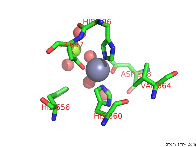
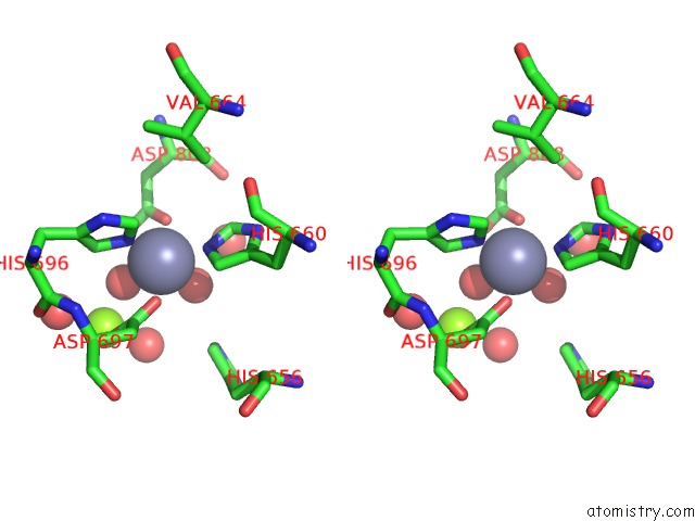
Zinc binding site 1 out of 4 in 4htx
Go back to
Zinc binding site 1 out
of 4 in the Crystal Structure of PDE2 Catalytic Domain in Complex with BAY60-7550
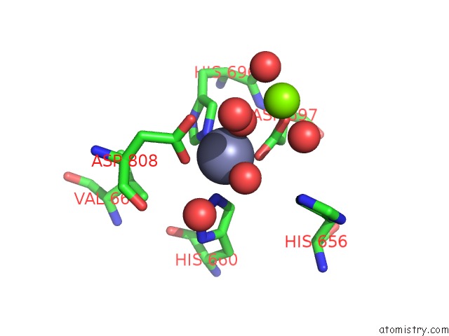
Mono view
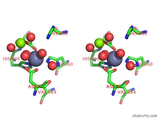
Stereo pair view
Mono view
Stereo pair view
A full contact list of Zinc with other atoms in the Zn binding
site number 1 of Crystal Structure of PDE2 Catalytic Domain in Complex with BAY60-7550 within 5.0Å range:
|
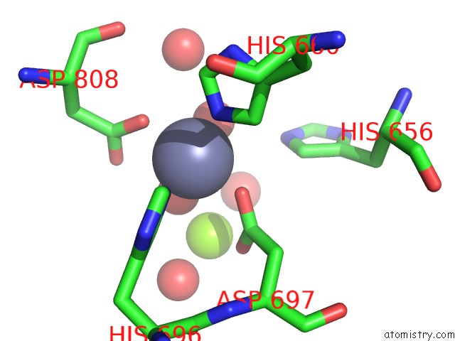
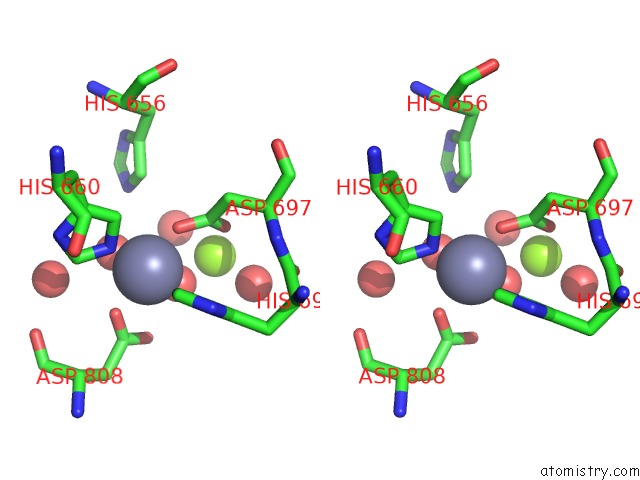
Zinc binding site 2 out of 4 in 4htx
Go back to
Zinc binding site 2 out
of 4 in the Crystal Structure of PDE2 Catalytic Domain in Complex with BAY60-7550
Mono view
Stereo pair view
Mono view
Stereo pair view
A full contact list of Zinc with other atoms in the Zn binding
site number 2 of Crystal Structure of PDE2 Catalytic Domain in Complex with BAY60-7550 within 5.0Å range:
|
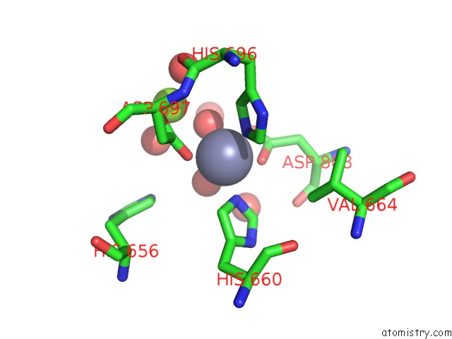
Zinc binding site 3 out of 4 in 4htx
Go back to
Zinc binding site 3 out
of 4 in the Crystal Structure of PDE2 Catalytic Domain in Complex with BAY60-7550
Mono view
Stereo pair view
Mono view
Stereo pair view
A full contact list of Zinc with other atoms in the Zn binding
site number 3 of Crystal Structure of PDE2 Catalytic Domain in Complex with BAY60-7550 within 5.0Å range:
|
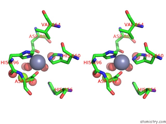
Zinc binding site 4 out of 4 in 4htx
Go back to
Zinc binding site 4 out
of 4 in the Crystal Structure of PDE2 Catalytic Domain in Complex with BAY60-7550
Mono view
Stereo pair view
Mono view
Stereo pair view
A full contact list of Zinc with other atoms in the Zn binding
site number 4 of Crystal Structure of PDE2 Catalytic Domain in Complex with BAY60-7550 within 5.0Å range:
|
Reference:
J.Zhu,
Q.Yang,
D.Dai,
Q.Huang.
X-Ray Crystal Structure of Phosphodiesterase 2 in Complex with A Highly Selective, Nanomolar Inhibitor Reveals A Binding-Induced Pocket Important For Selectivity. J.Am.Chem.Soc. V. 135 11708 2013.
ISSN: ISSN 0002-7863
PubMed: 23899287
DOI: 10.1021/JA404449G
Page generated: Sun Oct 27 00:17:39 2024
ISSN: ISSN 0002-7863
PubMed: 23899287
DOI: 10.1021/JA404449G
Last articles
Zn in 9JYWZn in 9IR4
Zn in 9IR3
Zn in 9GMX
Zn in 9GMW
Zn in 9JEJ
Zn in 9ERF
Zn in 9ERE
Zn in 9EGV
Zn in 9EGW