Zinc »
PDB 4h8p-4hk6 »
4hcc »
Zinc in PDB 4hcc: The Zinc Ion Bound Form of Crystal Structure of E.Coli Exoi-Ssdna Complex
Enzymatic activity of The Zinc Ion Bound Form of Crystal Structure of E.Coli Exoi-Ssdna Complex
All present enzymatic activity of The Zinc Ion Bound Form of Crystal Structure of E.Coli Exoi-Ssdna Complex:
3.1.11.1;
3.1.11.1;
Protein crystallography data
The structure of The Zinc Ion Bound Form of Crystal Structure of E.Coli Exoi-Ssdna Complex, PDB code: 4hcc
was solved by
R.Qiu,
J.Wei,
T.Lou,
M.Liu,
C.Ji,
W.Gong,
with X-Ray Crystallography technique. A brief refinement statistics is given in the table below:
| Resolution Low / High (Å) | 30.00 / 2.96 |
| Space group | P 21 21 21 |
| Cell size a, b, c (Å), α, β, γ (°) | 63.037, 108.692, 158.403, 90.00, 90.00, 90.00 |
| R / Rfree (%) | 18.5 / 24.5 |
Zinc Binding Sites:
The binding sites of Zinc atom in the The Zinc Ion Bound Form of Crystal Structure of E.Coli Exoi-Ssdna Complex
(pdb code 4hcc). This binding sites where shown within
5.0 Angstroms radius around Zinc atom.
In total 2 binding sites of Zinc where determined in the The Zinc Ion Bound Form of Crystal Structure of E.Coli Exoi-Ssdna Complex, PDB code: 4hcc:
Jump to Zinc binding site number: 1; 2;
In total 2 binding sites of Zinc where determined in the The Zinc Ion Bound Form of Crystal Structure of E.Coli Exoi-Ssdna Complex, PDB code: 4hcc:
Jump to Zinc binding site number: 1; 2;
Zinc binding site 1 out of 2 in 4hcc
Go back to
Zinc binding site 1 out
of 2 in the The Zinc Ion Bound Form of Crystal Structure of E.Coli Exoi-Ssdna Complex
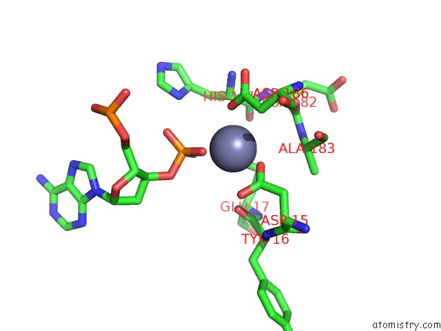
Mono view
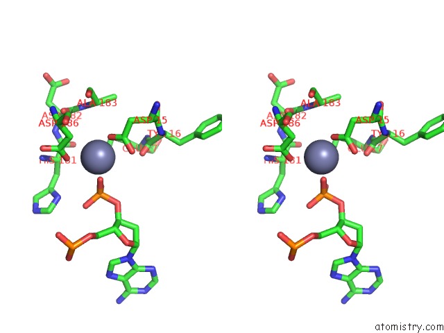
Stereo pair view
Mono view
Stereo pair view
A full contact list of Zinc with other atoms in the Zn binding
site number 1 of The Zinc Ion Bound Form of Crystal Structure of E.Coli Exoi-Ssdna Complex within 5.0Å range:
|
Zinc binding site 2 out of 2 in 4hcc
Go back to
Zinc binding site 2 out
of 2 in the The Zinc Ion Bound Form of Crystal Structure of E.Coli Exoi-Ssdna Complex
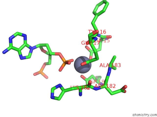
Mono view
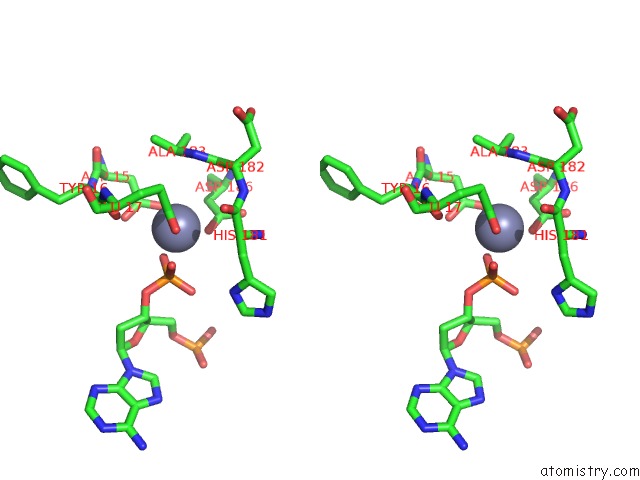
Stereo pair view
Mono view
Stereo pair view
A full contact list of Zinc with other atoms in the Zn binding
site number 2 of The Zinc Ion Bound Form of Crystal Structure of E.Coli Exoi-Ssdna Complex within 5.0Å range:
|
Reference:
R.Qiu,
T.Lou,
J.Wei,
M.Liu,
S.Gu,
R.Tang,
C.Ji,
W.Gong.
The Structures of Escherichia Coli Exonuclease I in Complex with the Single Strand Dna To Be Published.
Page generated: Sun Oct 27 00:00:03 2024
Last articles
Zn in 9JYWZn in 9IR4
Zn in 9IR3
Zn in 9GMX
Zn in 9GMW
Zn in 9JEJ
Zn in 9ERF
Zn in 9ERE
Zn in 9EGV
Zn in 9EGW