Zinc »
PDB 4gy1-4h84 »
4h0f »
Zinc in PDB 4h0f: Mutant Structure of Laminin-Binding Adhesin (Lmb) From Streptococcus Agalactiae
Protein crystallography data
The structure of Mutant Structure of Laminin-Binding Adhesin (Lmb) From Streptococcus Agalactiae, PDB code: 4h0f
was solved by
P.Karthe,
R.Preethi,
with X-Ray Crystallography technique. A brief refinement statistics is given in the table below:
| Resolution Low / High (Å) | 20.00 / 2.40 |
| Space group | P 1 21 1 |
| Cell size a, b, c (Å), α, β, γ (°) | 42.400, 93.990, 66.880, 90.00, 105.42, 90.00 |
| R / Rfree (%) | 24 / 28.1 |
Zinc Binding Sites:
The binding sites of Zinc atom in the Mutant Structure of Laminin-Binding Adhesin (Lmb) From Streptococcus Agalactiae
(pdb code 4h0f). This binding sites where shown within
5.0 Angstroms radius around Zinc atom.
In total 2 binding sites of Zinc where determined in the Mutant Structure of Laminin-Binding Adhesin (Lmb) From Streptococcus Agalactiae, PDB code: 4h0f:
Jump to Zinc binding site number: 1; 2;
In total 2 binding sites of Zinc where determined in the Mutant Structure of Laminin-Binding Adhesin (Lmb) From Streptococcus Agalactiae, PDB code: 4h0f:
Jump to Zinc binding site number: 1; 2;
Zinc binding site 1 out of 2 in 4h0f
Go back to
Zinc binding site 1 out
of 2 in the Mutant Structure of Laminin-Binding Adhesin (Lmb) From Streptococcus Agalactiae
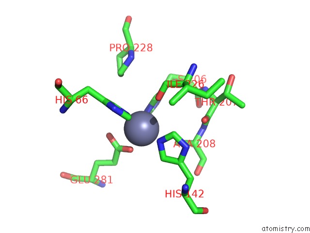
Mono view
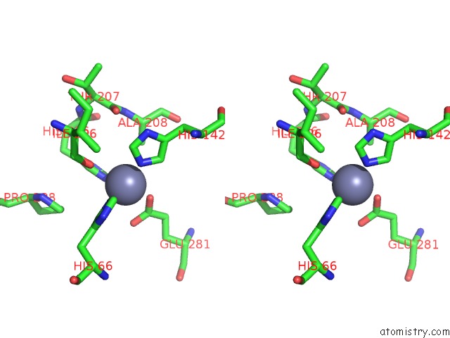
Stereo pair view
Mono view
Stereo pair view
A full contact list of Zinc with other atoms in the Zn binding
site number 1 of Mutant Structure of Laminin-Binding Adhesin (Lmb) From Streptococcus Agalactiae within 5.0Å range:
|
Zinc binding site 2 out of 2 in 4h0f
Go back to
Zinc binding site 2 out
of 2 in the Mutant Structure of Laminin-Binding Adhesin (Lmb) From Streptococcus Agalactiae
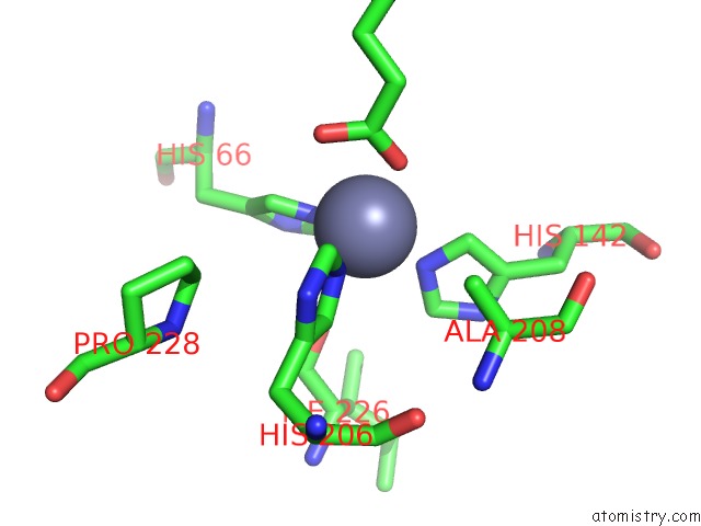
Mono view
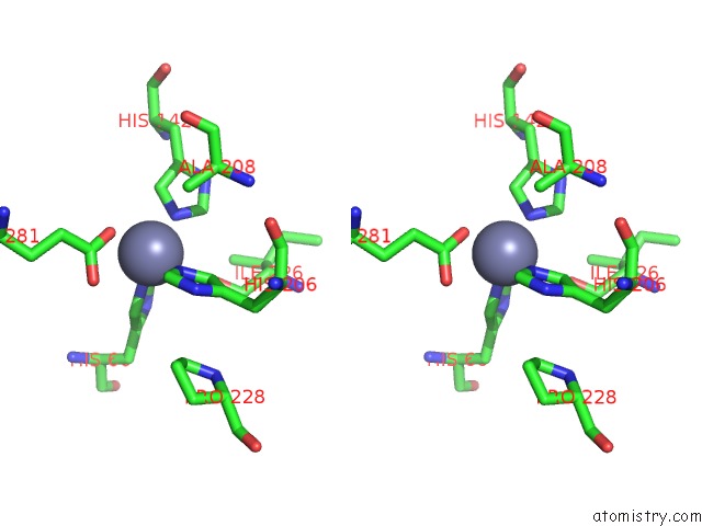
Stereo pair view
Mono view
Stereo pair view
A full contact list of Zinc with other atoms in the Zn binding
site number 2 of Mutant Structure of Laminin-Binding Adhesin (Lmb) From Streptococcus Agalactiae within 5.0Å range:
|
Reference:
P.Ragunathan,
D.Sridaran,
A.Weigel,
S.Shabayek,
B.Spellerberg,
K.Ponnuraj.
Metal Binding Is Critical For the Folding and Function of Laminin Binding Protein, Lmb of Streptococcus Agalactiae. Plos One V. 8 67517 2013.
ISSN: ESSN 1932-6203
PubMed: 23826314
DOI: 10.1371/JOURNAL.PONE.0067517
Page generated: Sat Oct 26 23:46:18 2024
ISSN: ESSN 1932-6203
PubMed: 23826314
DOI: 10.1371/JOURNAL.PONE.0067517
Last articles
Zn in 9JYWZn in 9IR4
Zn in 9IR3
Zn in 9GMX
Zn in 9GMW
Zn in 9JEJ
Zn in 9ERF
Zn in 9ERE
Zn in 9EGV
Zn in 9EGW