Zinc »
PDB 4eez-4eyf »
4eyb »
Zinc in PDB 4eyb: Crystal Structure of Ndm-1 Bound to Hydrolyzed Oxacillin
Enzymatic activity of Crystal Structure of Ndm-1 Bound to Hydrolyzed Oxacillin
All present enzymatic activity of Crystal Structure of Ndm-1 Bound to Hydrolyzed Oxacillin:
3.5.2.6;
3.5.2.6;
Protein crystallography data
The structure of Crystal Structure of Ndm-1 Bound to Hydrolyzed Oxacillin, PDB code: 4eyb
was solved by
N.C.J.Strynadka,
D.T.King,
with X-Ray Crystallography technique. A brief refinement statistics is given in the table below:
| Resolution Low / High (Å) | 25.42 / 1.16 |
| Space group | P 21 21 21 |
| Cell size a, b, c (Å), α, β, γ (°) | 39.200, 79.370, 134.150, 90.00, 90.00, 90.00 |
| R / Rfree (%) | 13.5 / 16.4 |
Zinc Binding Sites:
The binding sites of Zinc atom in the Crystal Structure of Ndm-1 Bound to Hydrolyzed Oxacillin
(pdb code 4eyb). This binding sites where shown within
5.0 Angstroms radius around Zinc atom.
In total 5 binding sites of Zinc where determined in the Crystal Structure of Ndm-1 Bound to Hydrolyzed Oxacillin, PDB code: 4eyb:
Jump to Zinc binding site number: 1; 2; 3; 4; 5;
In total 5 binding sites of Zinc where determined in the Crystal Structure of Ndm-1 Bound to Hydrolyzed Oxacillin, PDB code: 4eyb:
Jump to Zinc binding site number: 1; 2; 3; 4; 5;
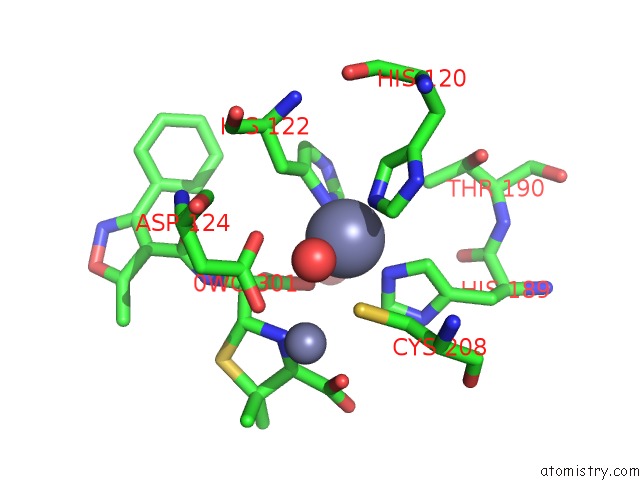
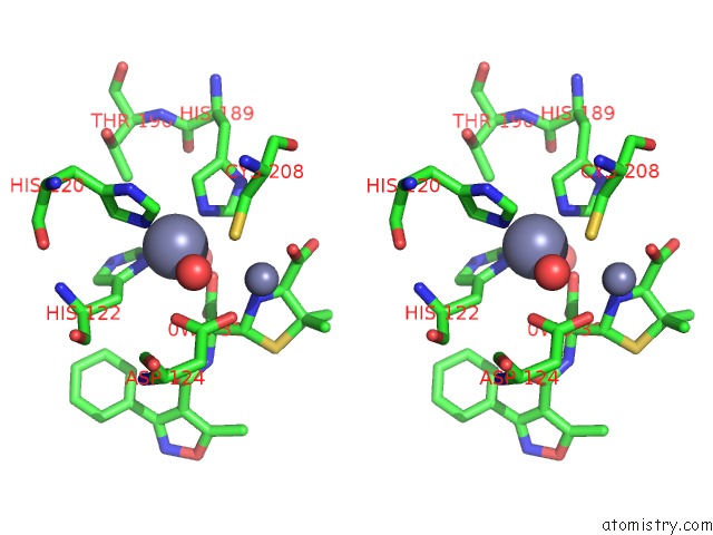
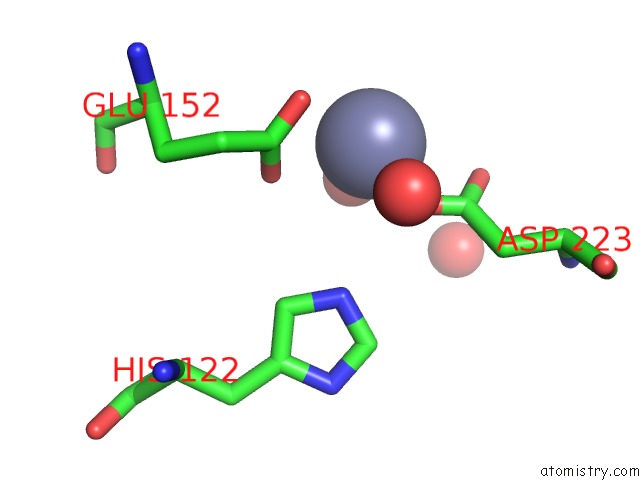
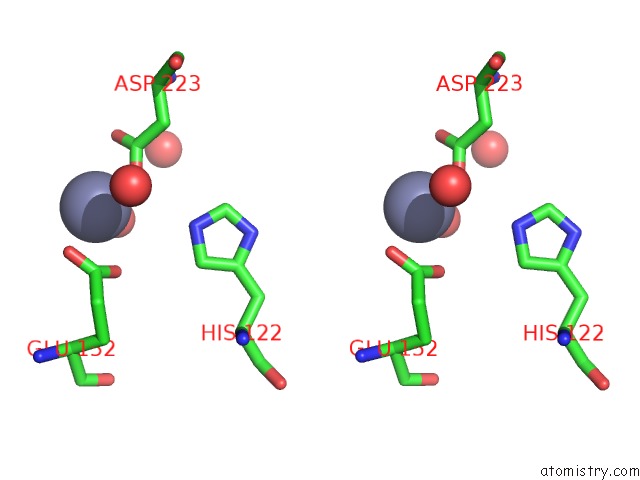
Zinc binding site 1 out of 5 in 4eyb
Go back to
Zinc binding site 1 out
of 5 in the Crystal Structure of Ndm-1 Bound to Hydrolyzed Oxacillin

Mono view

Stereo pair view
Mono view
Stereo pair view
A full contact list of Zinc with other atoms in the Zn binding
site number 1 of Crystal Structure of Ndm-1 Bound to Hydrolyzed Oxacillin within 5.0Å range:
|
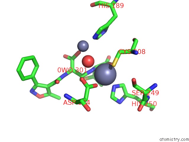
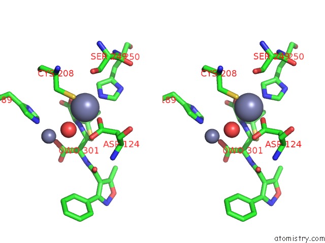
Zinc binding site 2 out of 5 in 4eyb
Go back to
Zinc binding site 2 out
of 5 in the Crystal Structure of Ndm-1 Bound to Hydrolyzed Oxacillin

Mono view

Stereo pair view
Mono view
Stereo pair view
A full contact list of Zinc with other atoms in the Zn binding
site number 2 of Crystal Structure of Ndm-1 Bound to Hydrolyzed Oxacillin within 5.0Å range:
|
Zinc binding site 3 out of 5 in 4eyb
Go back to
Zinc binding site 3 out
of 5 in the Crystal Structure of Ndm-1 Bound to Hydrolyzed Oxacillin
Mono view
Stereo pair view
Mono view
Stereo pair view
A full contact list of Zinc with other atoms in the Zn binding
site number 3 of Crystal Structure of Ndm-1 Bound to Hydrolyzed Oxacillin within 5.0Å range:
|
Zinc binding site 4 out of 5 in 4eyb
Go back to
Zinc binding site 4 out
of 5 in the Crystal Structure of Ndm-1 Bound to Hydrolyzed Oxacillin
Mono view
Stereo pair view
Mono view
Stereo pair view
A full contact list of Zinc with other atoms in the Zn binding
site number 4 of Crystal Structure of Ndm-1 Bound to Hydrolyzed Oxacillin within 5.0Å range:
|
Zinc binding site 5 out of 5 in 4eyb
Go back to
Zinc binding site 5 out
of 5 in the Crystal Structure of Ndm-1 Bound to Hydrolyzed Oxacillin
Mono view
Stereo pair view
Mono view
Stereo pair view
A full contact list of Zinc with other atoms in the Zn binding
site number 5 of Crystal Structure of Ndm-1 Bound to Hydrolyzed Oxacillin within 5.0Å range:
|
Reference:
D.T.King,
L.J.Worrall,
R.Gruninger,
N.C.Strynadka.
New Delhi Metallo-Beta-Lactamase: Structural Insights Into Beta-Lactam Recognition and Inhibition J.Am.Chem.Soc. V. 134 11362 2012.
ISSN: ISSN 0002-7863
PubMed: 22713171
DOI: 10.1021/JA303579D
Page generated: Sat Oct 26 22:09:17 2024
ISSN: ISSN 0002-7863
PubMed: 22713171
DOI: 10.1021/JA303579D
Last articles
Zn in 9JYWZn in 9IR4
Zn in 9IR3
Zn in 9GMX
Zn in 9GMW
Zn in 9JEJ
Zn in 9ERF
Zn in 9ERE
Zn in 9EGV
Zn in 9EGW