Zinc »
PDB 4ad9-4arf »
4aqi »
Zinc in PDB 4aqi: Structure of Human S100A15 Bound to Zinc and Calcium
Protein crystallography data
The structure of Structure of Human S100A15 Bound to Zinc and Calcium, PDB code: 4aqi
was solved by
J.I.Murray,
M.L.Tonkin,
A.L.Whiting,
F.Peng,
B.Farnell,
F.Hof,
M.J.Boulanger,
with X-Ray Crystallography technique. A brief refinement statistics is given in the table below:
| Resolution Low / High (Å) | 28.15 / 1.70 |
| Space group | I 1 2 1 |
| Cell size a, b, c (Å), α, β, γ (°) | 52.030, 33.470, 64.052, 90.00, 90.81, 90.00 |
| R / Rfree (%) | 19.808 / 23.045 |
Other elements in 4aqi:
The structure of Structure of Human S100A15 Bound to Zinc and Calcium also contains other interesting chemical elements:
| Chlorine | (Cl) | 1 atom |
| Calcium | (Ca) | 1 atom |
Zinc Binding Sites:
The binding sites of Zinc atom in the Structure of Human S100A15 Bound to Zinc and Calcium
(pdb code 4aqi). This binding sites where shown within
5.0 Angstroms radius around Zinc atom.
In total 2 binding sites of Zinc where determined in the Structure of Human S100A15 Bound to Zinc and Calcium, PDB code: 4aqi:
Jump to Zinc binding site number: 1; 2;
In total 2 binding sites of Zinc where determined in the Structure of Human S100A15 Bound to Zinc and Calcium, PDB code: 4aqi:
Jump to Zinc binding site number: 1; 2;
Zinc binding site 1 out of 2 in 4aqi
Go back to
Zinc binding site 1 out
of 2 in the Structure of Human S100A15 Bound to Zinc and Calcium
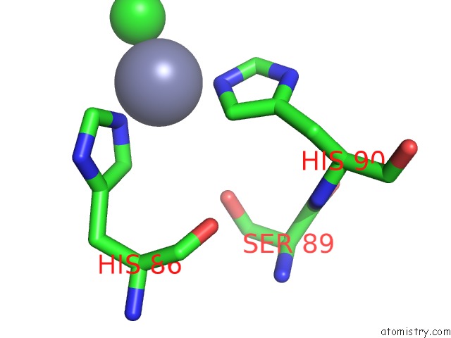
Mono view
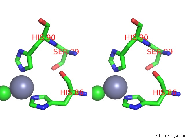
Stereo pair view
Mono view
Stereo pair view
A full contact list of Zinc with other atoms in the Zn binding
site number 1 of Structure of Human S100A15 Bound to Zinc and Calcium within 5.0Å range:
|
Zinc binding site 2 out of 2 in 4aqi
Go back to
Zinc binding site 2 out
of 2 in the Structure of Human S100A15 Bound to Zinc and Calcium
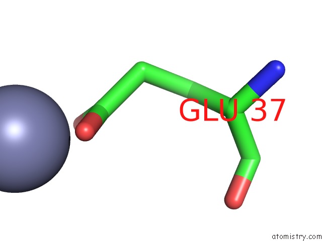
Mono view
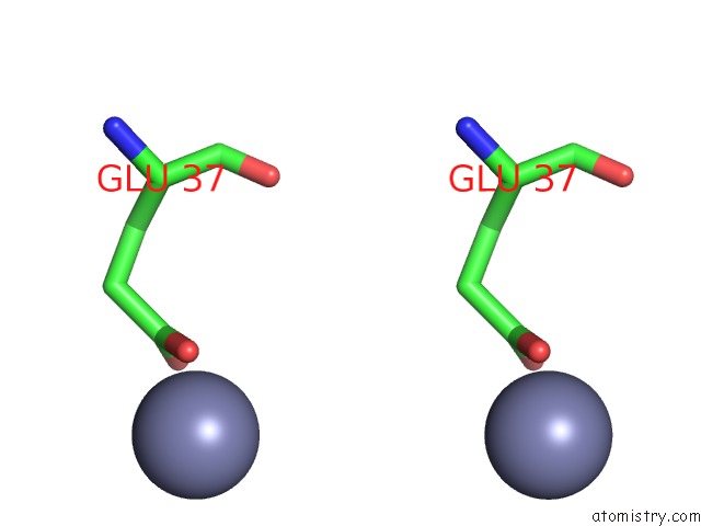
Stereo pair view
Mono view
Stereo pair view
A full contact list of Zinc with other atoms in the Zn binding
site number 2 of Structure of Human S100A15 Bound to Zinc and Calcium within 5.0Å range:
|
Reference:
J.I.Murray,
M.L.Tonkin,
A.L.Whiting,
F.Peng,
B.Farnell,
J.T.Cullen,
F.Hof,
M.J.Boulanger.
Structural Characterization of S100A15 Reveals A Novel Zinc Coordination Site Among S100 Proteins and Altered Surface Chemistry with Functional Implications For Receptor Binding. Bmc Struct.Biol. V. 12 16 2012.
ISSN: ISSN 1472-6807
PubMed: 22747601
DOI: 10.1186/1472-6807-12-16
Page generated: Sat Oct 26 19:19:19 2024
ISSN: ISSN 1472-6807
PubMed: 22747601
DOI: 10.1186/1472-6807-12-16
Last articles
Zn in 9MJ5Zn in 9HNW
Zn in 9G0L
Zn in 9FNE
Zn in 9DZN
Zn in 9E0I
Zn in 9D32
Zn in 9DAK
Zn in 8ZXC
Zn in 8ZUF