Zinc »
PDB 4a3f-4aa6 »
4aa1 »
Zinc in PDB 4aa1: Crystal Structure of Ance in Complex with Angiotensin-II
Enzymatic activity of Crystal Structure of Ance in Complex with Angiotensin-II
All present enzymatic activity of Crystal Structure of Ance in Complex with Angiotensin-II:
3.4.15.1;
3.4.15.1;
Protein crystallography data
The structure of Crystal Structure of Ance in Complex with Angiotensin-II, PDB code: 4aa1
was solved by
R.E.Isaac,
M.Akif,
S.L.U.Schwager,
G.Masuyer,
E.D.Sturrock,
K.R.Acharya,
with X-Ray Crystallography technique. A brief refinement statistics is given in the table below:
| Resolution Low / High (Å) | 38.57 / 1.99 |
| Space group | H 3 |
| Cell size a, b, c (Å), α, β, γ (°) | 173.414, 173.414, 102.245, 90.00, 90.00, 120.00 |
| R / Rfree (%) | 18.835 / 21.072 |
Zinc Binding Sites:
The binding sites of Zinc atom in the Crystal Structure of Ance in Complex with Angiotensin-II
(pdb code 4aa1). This binding sites where shown within
5.0 Angstroms radius around Zinc atom.
In total only one binding site of Zinc was determined in the Crystal Structure of Ance in Complex with Angiotensin-II, PDB code: 4aa1:
In total only one binding site of Zinc was determined in the Crystal Structure of Ance in Complex with Angiotensin-II, PDB code: 4aa1:
Zinc binding site 1 out of 1 in 4aa1
Go back to
Zinc binding site 1 out
of 1 in the Crystal Structure of Ance in Complex with Angiotensin-II
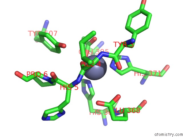
Mono view
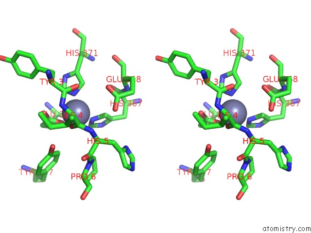
Stereo pair view
Mono view
Stereo pair view
A full contact list of Zinc with other atoms in the Zn binding
site number 1 of Crystal Structure of Ance in Complex with Angiotensin-II within 5.0Å range:
|
Reference:
M.Akif,
G.Masuyer,
R.J.Bingham,
E.D.Sturrock,
R.E.Isaac,
K.R.Acharya.
Structural Basis of Peptide Recognition By the Angiotensin-I Converting Enzyme Homologue Ance From Drosophila Melanogaster Febs J. V. 279 4525 2012.
ISSN: ISSN 1742-464X
PubMed: 23082758
DOI: 10.1111/FEBS.12038
Page generated: Sat Oct 26 19:08:13 2024
ISSN: ISSN 1742-464X
PubMed: 23082758
DOI: 10.1111/FEBS.12038
Last articles
Zn in 9JYWZn in 9IR4
Zn in 9IR3
Zn in 9GMX
Zn in 9GMW
Zn in 9JEJ
Zn in 9ERF
Zn in 9ERE
Zn in 9EGV
Zn in 9EGW