Zinc »
PDB 3w52-3wnq »
3wlf »
Zinc in PDB 3wlf: Crystal Structure of (R)-Carbonyl Reductase From Candida Parapsilosis in Complex with (R)-1-Phenyl-1,2-Ethanediol
Enzymatic activity of Crystal Structure of (R)-Carbonyl Reductase From Candida Parapsilosis in Complex with (R)-1-Phenyl-1,2-Ethanediol
All present enzymatic activity of Crystal Structure of (R)-Carbonyl Reductase From Candida Parapsilosis in Complex with (R)-1-Phenyl-1,2-Ethanediol:
1.1.1.1;
1.1.1.1;
Protein crystallography data
The structure of Crystal Structure of (R)-Carbonyl Reductase From Candida Parapsilosis in Complex with (R)-1-Phenyl-1,2-Ethanediol, PDB code: 3wlf
was solved by
S.S.Wang,
Y.Nie,
Y.Xu,
R.Z.Zhang,
C.H.Huang,
H.C.Chan,
R.T.Guo,
R.Xiao,
with X-Ray Crystallography technique. A brief refinement statistics is given in the table below:
| Resolution Low / High (Å) | 25.00 / 2.30 |
| Space group | P 21 21 21 |
| Cell size a, b, c (Å), α, β, γ (°) | 93.493, 103.722, 141.941, 90.00, 90.00, 90.00 |
| R / Rfree (%) | 20.6 / 25 |
Zinc Binding Sites:
The binding sites of Zinc atom in the Crystal Structure of (R)-Carbonyl Reductase From Candida Parapsilosis in Complex with (R)-1-Phenyl-1,2-Ethanediol
(pdb code 3wlf). This binding sites where shown within
5.0 Angstroms radius around Zinc atom.
In total 8 binding sites of Zinc where determined in the Crystal Structure of (R)-Carbonyl Reductase From Candida Parapsilosis in Complex with (R)-1-Phenyl-1,2-Ethanediol, PDB code: 3wlf:
Jump to Zinc binding site number: 1; 2; 3; 4; 5; 6; 7; 8;
In total 8 binding sites of Zinc where determined in the Crystal Structure of (R)-Carbonyl Reductase From Candida Parapsilosis in Complex with (R)-1-Phenyl-1,2-Ethanediol, PDB code: 3wlf:
Jump to Zinc binding site number: 1; 2; 3; 4; 5; 6; 7; 8;
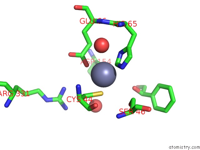
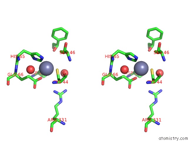
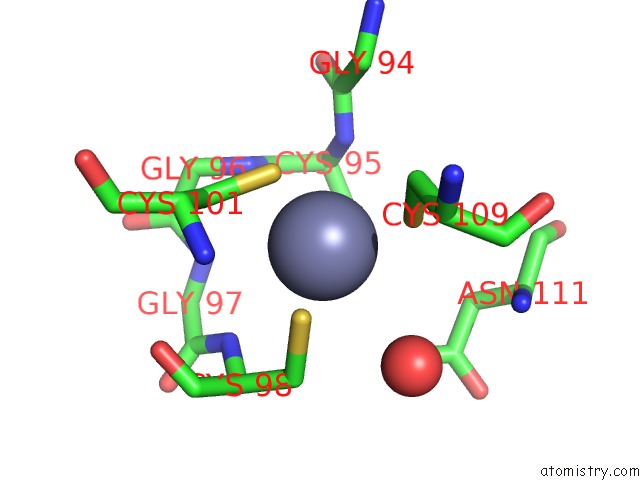
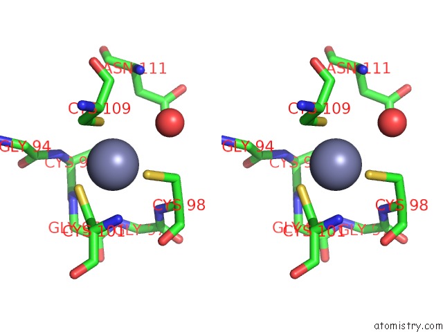
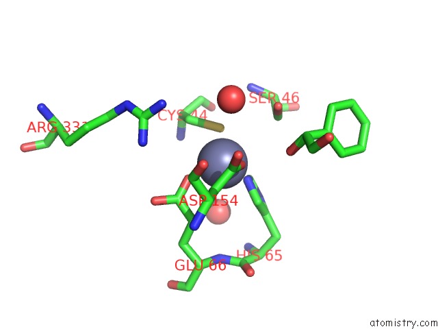
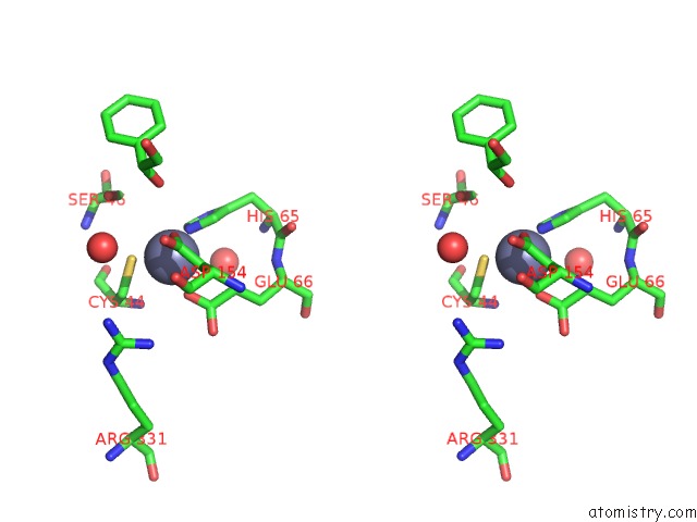
Zinc binding site 1 out of 8 in 3wlf
Go back to
Zinc binding site 1 out
of 8 in the Crystal Structure of (R)-Carbonyl Reductase From Candida Parapsilosis in Complex with (R)-1-Phenyl-1,2-Ethanediol
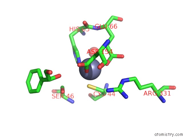
Mono view
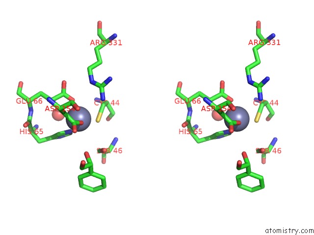
Stereo pair view
Mono view
Stereo pair view
A full contact list of Zinc with other atoms in the Zn binding
site number 1 of Crystal Structure of (R)-Carbonyl Reductase From Candida Parapsilosis in Complex with (R)-1-Phenyl-1,2-Ethanediol within 5.0Å range:
|
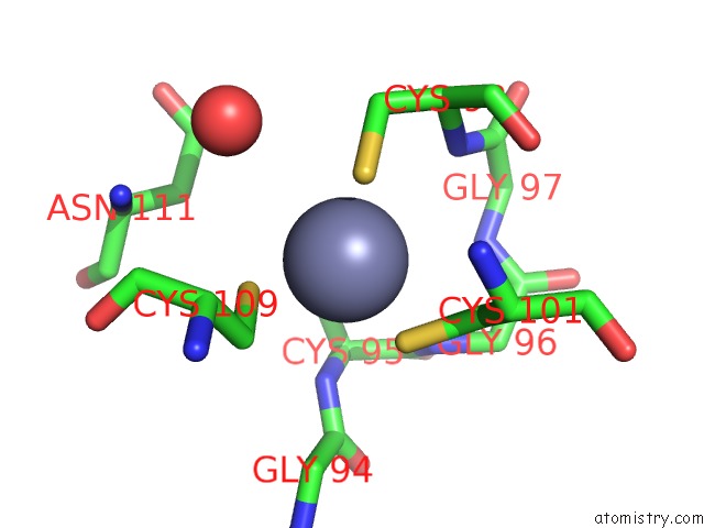
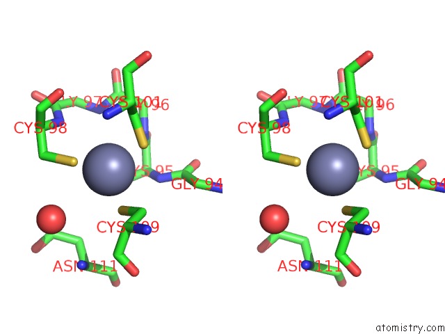
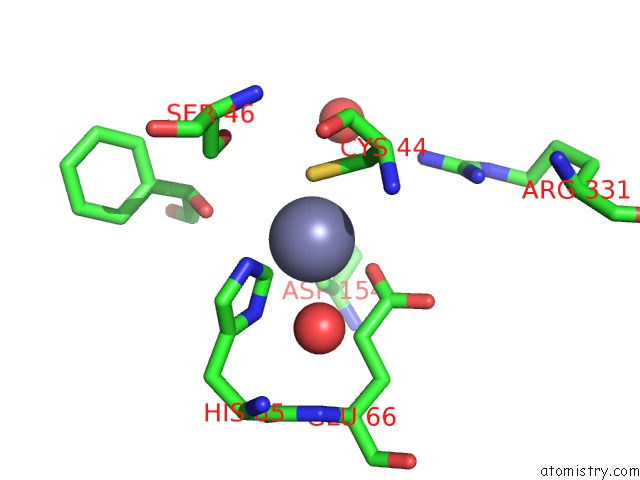
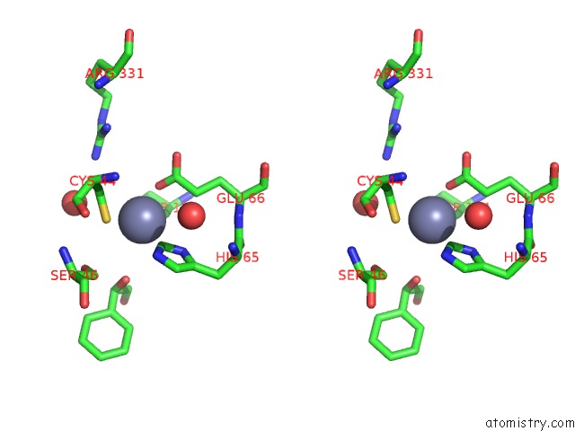
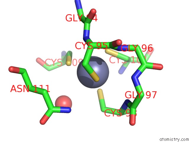
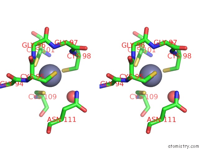
Zinc binding site 2 out of 8 in 3wlf
Go back to
Zinc binding site 2 out
of 8 in the Crystal Structure of (R)-Carbonyl Reductase From Candida Parapsilosis in Complex with (R)-1-Phenyl-1,2-Ethanediol
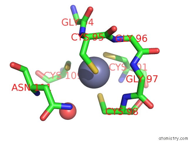
Mono view
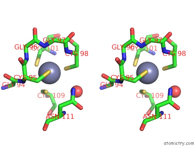
Stereo pair view
Mono view
Stereo pair view
A full contact list of Zinc with other atoms in the Zn binding
site number 2 of Crystal Structure of (R)-Carbonyl Reductase From Candida Parapsilosis in Complex with (R)-1-Phenyl-1,2-Ethanediol within 5.0Å range:
|
Zinc binding site 3 out of 8 in 3wlf
Go back to
Zinc binding site 3 out
of 8 in the Crystal Structure of (R)-Carbonyl Reductase From Candida Parapsilosis in Complex with (R)-1-Phenyl-1,2-Ethanediol
Mono view
Stereo pair view
Mono view
Stereo pair view
A full contact list of Zinc with other atoms in the Zn binding
site number 3 of Crystal Structure of (R)-Carbonyl Reductase From Candida Parapsilosis in Complex with (R)-1-Phenyl-1,2-Ethanediol within 5.0Å range:
|
Zinc binding site 4 out of 8 in 3wlf
Go back to
Zinc binding site 4 out
of 8 in the Crystal Structure of (R)-Carbonyl Reductase From Candida Parapsilosis in Complex with (R)-1-Phenyl-1,2-Ethanediol
Mono view
Stereo pair view
Mono view
Stereo pair view
A full contact list of Zinc with other atoms in the Zn binding
site number 4 of Crystal Structure of (R)-Carbonyl Reductase From Candida Parapsilosis in Complex with (R)-1-Phenyl-1,2-Ethanediol within 5.0Å range:
|
Zinc binding site 5 out of 8 in 3wlf
Go back to
Zinc binding site 5 out
of 8 in the Crystal Structure of (R)-Carbonyl Reductase From Candida Parapsilosis in Complex with (R)-1-Phenyl-1,2-Ethanediol
Mono view
Stereo pair view
Mono view
Stereo pair view
A full contact list of Zinc with other atoms in the Zn binding
site number 5 of Crystal Structure of (R)-Carbonyl Reductase From Candida Parapsilosis in Complex with (R)-1-Phenyl-1,2-Ethanediol within 5.0Å range:
|
Zinc binding site 6 out of 8 in 3wlf
Go back to
Zinc binding site 6 out
of 8 in the Crystal Structure of (R)-Carbonyl Reductase From Candida Parapsilosis in Complex with (R)-1-Phenyl-1,2-Ethanediol
Mono view
Stereo pair view
Mono view
Stereo pair view
A full contact list of Zinc with other atoms in the Zn binding
site number 6 of Crystal Structure of (R)-Carbonyl Reductase From Candida Parapsilosis in Complex with (R)-1-Phenyl-1,2-Ethanediol within 5.0Å range:
|
Zinc binding site 7 out of 8 in 3wlf
Go back to
Zinc binding site 7 out
of 8 in the Crystal Structure of (R)-Carbonyl Reductase From Candida Parapsilosis in Complex with (R)-1-Phenyl-1,2-Ethanediol
Mono view
Stereo pair view
Mono view
Stereo pair view
A full contact list of Zinc with other atoms in the Zn binding
site number 7 of Crystal Structure of (R)-Carbonyl Reductase From Candida Parapsilosis in Complex with (R)-1-Phenyl-1,2-Ethanediol within 5.0Å range:
|
Zinc binding site 8 out of 8 in 3wlf
Go back to
Zinc binding site 8 out
of 8 in the Crystal Structure of (R)-Carbonyl Reductase From Candida Parapsilosis in Complex with (R)-1-Phenyl-1,2-Ethanediol
Mono view
Stereo pair view
Mono view
Stereo pair view
A full contact list of Zinc with other atoms in the Zn binding
site number 8 of Crystal Structure of (R)-Carbonyl Reductase From Candida Parapsilosis in Complex with (R)-1-Phenyl-1,2-Ethanediol within 5.0Å range:
|
Reference:
S.S.Wang,
Y.Nie,
Y.Xu,
R.Z.Zhang,
T.P.Ko,
C.H.Huang,
H.C.Chan,
R.T.Guo,
R.Xiao.
Unconserved Substrate-Binding Sites Direct the Stereoselectivity of Medium-Chain Alcohol Dehydrogenase Chem.Commun.(Camb.) V. 50 7770 2014.
ISSN: ISSN 1359-7345
PubMed: 24834985
DOI: 10.1039/C4CC01752H
Page generated: Sat Oct 26 18:08:06 2024
ISSN: ISSN 1359-7345
PubMed: 24834985
DOI: 10.1039/C4CC01752H
Last articles
Zn in 9JYWZn in 9IR4
Zn in 9IR3
Zn in 9GMX
Zn in 9GMW
Zn in 9JEJ
Zn in 9ERF
Zn in 9ERE
Zn in 9EGV
Zn in 9EGW