Zinc »
PDB 3v2m-3vh1 »
3vgk »
Zinc in PDB 3vgk: Crystal Structure of A Rok Family Glucokinase From Streptomyces Griseus
Enzymatic activity of Crystal Structure of A Rok Family Glucokinase From Streptomyces Griseus
All present enzymatic activity of Crystal Structure of A Rok Family Glucokinase From Streptomyces Griseus:
2.7.1.2;
2.7.1.2;
Protein crystallography data
The structure of Crystal Structure of A Rok Family Glucokinase From Streptomyces Griseus, PDB code: 3vgk
was solved by
K.Miyazono,
N.Tabei,
S.Morita,
Y.Ohnishi,
S.Horinouchi,
M.Tanokura,
with X-Ray Crystallography technique. A brief refinement statistics is given in the table below:
| Resolution Low / High (Å) | 20.00 / 3.25 |
| Space group | P 1 21 1 |
| Cell size a, b, c (Å), α, β, γ (°) | 98.690, 173.700, 124.030, 90.00, 106.69, 90.00 |
| R / Rfree (%) | 20.6 / 26.4 |
Zinc Binding Sites:
The binding sites of Zinc atom in the Crystal Structure of A Rok Family Glucokinase From Streptomyces Griseus
(pdb code 3vgk). This binding sites where shown within
5.0 Angstroms radius around Zinc atom.
In total 8 binding sites of Zinc where determined in the Crystal Structure of A Rok Family Glucokinase From Streptomyces Griseus, PDB code: 3vgk:
Jump to Zinc binding site number: 1; 2; 3; 4; 5; 6; 7; 8;
In total 8 binding sites of Zinc where determined in the Crystal Structure of A Rok Family Glucokinase From Streptomyces Griseus, PDB code: 3vgk:
Jump to Zinc binding site number: 1; 2; 3; 4; 5; 6; 7; 8;
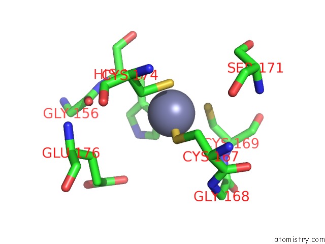
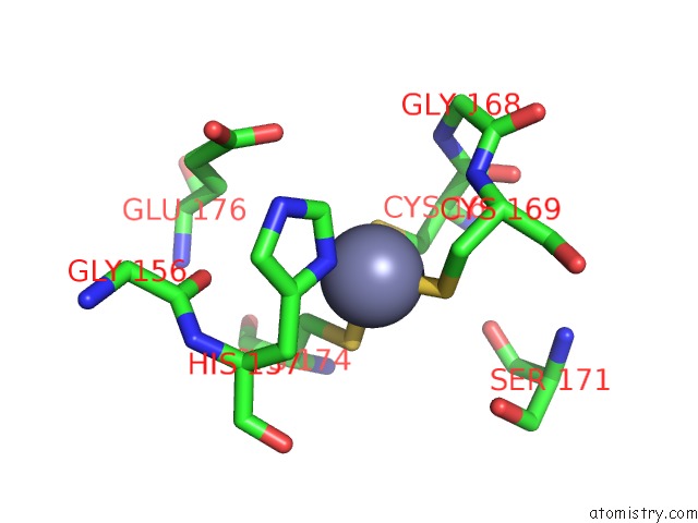
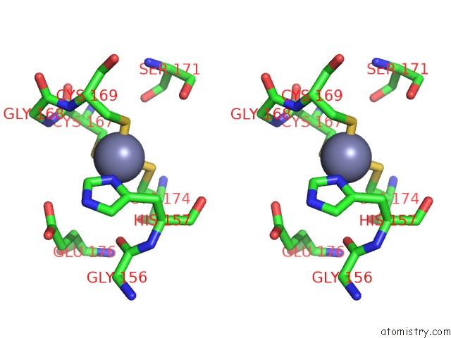
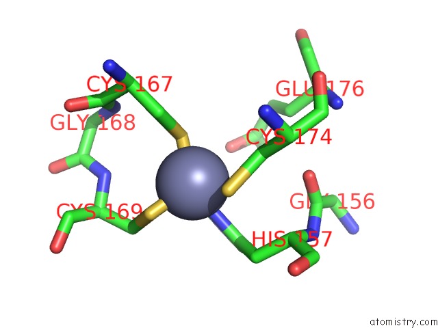
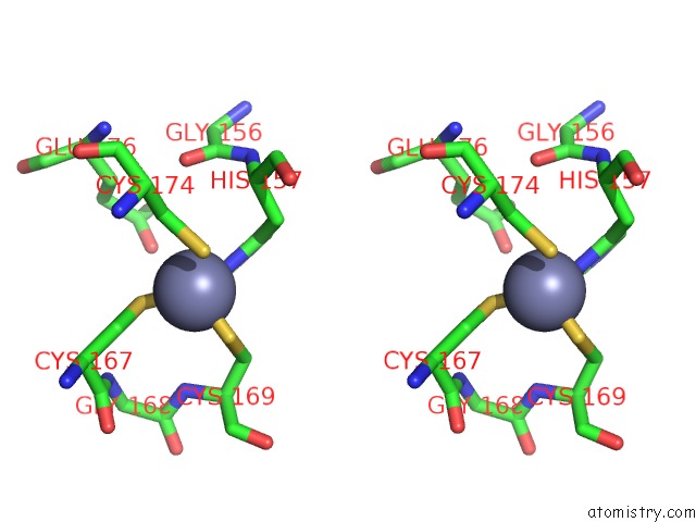
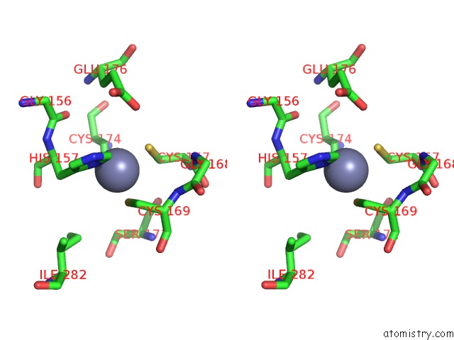
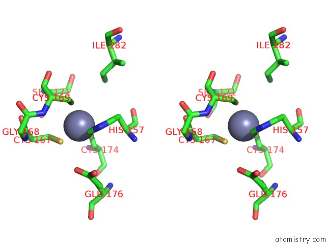
Zinc binding site 1 out of 8 in 3vgk
Go back to
Zinc binding site 1 out
of 8 in the Crystal Structure of A Rok Family Glucokinase From Streptomyces Griseus

Mono view
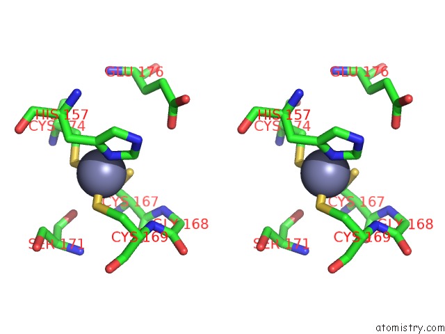
Stereo pair view
Mono view
Stereo pair view
A full contact list of Zinc with other atoms in the Zn binding
site number 1 of Crystal Structure of A Rok Family Glucokinase From Streptomyces Griseus within 5.0Å range:
|
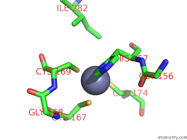
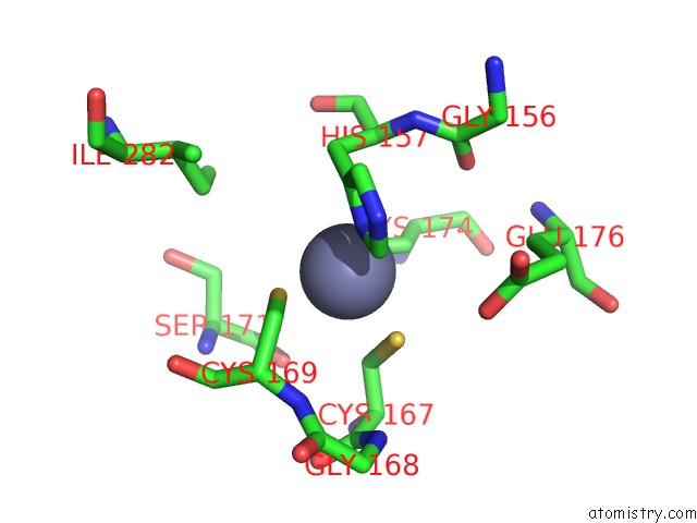
Zinc binding site 2 out of 8 in 3vgk
Go back to
Zinc binding site 2 out
of 8 in the Crystal Structure of A Rok Family Glucokinase From Streptomyces Griseus
Mono view

Stereo pair view
Mono view
Stereo pair view
A full contact list of Zinc with other atoms in the Zn binding
site number 2 of Crystal Structure of A Rok Family Glucokinase From Streptomyces Griseus within 5.0Å range:
|
Zinc binding site 3 out of 8 in 3vgk
Go back to
Zinc binding site 3 out
of 8 in the Crystal Structure of A Rok Family Glucokinase From Streptomyces Griseus
Mono view
Stereo pair view
Mono view
Stereo pair view
A full contact list of Zinc with other atoms in the Zn binding
site number 3 of Crystal Structure of A Rok Family Glucokinase From Streptomyces Griseus within 5.0Å range:
|
Zinc binding site 4 out of 8 in 3vgk
Go back to
Zinc binding site 4 out
of 8 in the Crystal Structure of A Rok Family Glucokinase From Streptomyces Griseus
Mono view
Stereo pair view
Mono view
Stereo pair view
A full contact list of Zinc with other atoms in the Zn binding
site number 4 of Crystal Structure of A Rok Family Glucokinase From Streptomyces Griseus within 5.0Å range:
|
Zinc binding site 5 out of 8 in 3vgk
Go back to
Zinc binding site 5 out
of 8 in the Crystal Structure of A Rok Family Glucokinase From Streptomyces Griseus
Mono view

Stereo pair view
Mono view
Stereo pair view
A full contact list of Zinc with other atoms in the Zn binding
site number 5 of Crystal Structure of A Rok Family Glucokinase From Streptomyces Griseus within 5.0Å range:
|
Zinc binding site 6 out of 8 in 3vgk
Go back to
Zinc binding site 6 out
of 8 in the Crystal Structure of A Rok Family Glucokinase From Streptomyces Griseus

Mono view

Stereo pair view
Mono view
Stereo pair view
A full contact list of Zinc with other atoms in the Zn binding
site number 6 of Crystal Structure of A Rok Family Glucokinase From Streptomyces Griseus within 5.0Å range:
|
Zinc binding site 7 out of 8 in 3vgk
Go back to
Zinc binding site 7 out
of 8 in the Crystal Structure of A Rok Family Glucokinase From Streptomyces Griseus
Mono view
Stereo pair view
Mono view
Stereo pair view
A full contact list of Zinc with other atoms in the Zn binding
site number 7 of Crystal Structure of A Rok Family Glucokinase From Streptomyces Griseus within 5.0Å range:
|
Zinc binding site 8 out of 8 in 3vgk
Go back to
Zinc binding site 8 out
of 8 in the Crystal Structure of A Rok Family Glucokinase From Streptomyces Griseus

Mono view
Stereo pair view
Mono view
Stereo pair view
A full contact list of Zinc with other atoms in the Zn binding
site number 8 of Crystal Structure of A Rok Family Glucokinase From Streptomyces Griseus within 5.0Å range:
|
Reference:
K.Miyazono,
N.Tabei,
S.Morita,
Y.Ohnishi,
S.Horinouchi,
M.Tanokura.
Substrate Recognition Mechanism and Substrate-Dependent Conformational Changes of An Rok Family Glucokinase From Streptomyces Griseus J.Bacteriol. V. 194 607 2012.
ISSN: ISSN 0021-9193
PubMed: 22101842
DOI: 10.1128/JB.06173-11
Page generated: Sat Oct 26 17:46:10 2024
ISSN: ISSN 0021-9193
PubMed: 22101842
DOI: 10.1128/JB.06173-11
Last articles
Zn in 9JYWZn in 9IR4
Zn in 9IR3
Zn in 9GMX
Zn in 9GMW
Zn in 9JEJ
Zn in 9ERF
Zn in 9ERE
Zn in 9EGV
Zn in 9EGW