Zinc »
PDB 3udz-3ujp »
3uik »
Zinc in PDB 3uik: Crystal Structure of Human Survivin Mutant K62Y/H80W in Complex with H3(1-10) Peptide
Protein crystallography data
The structure of Crystal Structure of Human Survivin Mutant K62Y/H80W in Complex with H3(1-10) Peptide, PDB code: 3uik
was solved by
J.Du,
D.J.Patel,
with X-Ray Crystallography technique. A brief refinement statistics is given in the table below:
| Resolution Low / High (Å) | 27.68 / 2.70 |
| Space group | C 1 2 1 |
| Cell size a, b, c (Å), α, β, γ (°) | 115.503, 71.389, 81.874, 90.00, 128.73, 90.00 |
| R / Rfree (%) | 21.9 / 28.1 |
Zinc Binding Sites:
The binding sites of Zinc atom in the Crystal Structure of Human Survivin Mutant K62Y/H80W in Complex with H3(1-10) Peptide
(pdb code 3uik). This binding sites where shown within
5.0 Angstroms radius around Zinc atom.
In total 2 binding sites of Zinc where determined in the Crystal Structure of Human Survivin Mutant K62Y/H80W in Complex with H3(1-10) Peptide, PDB code: 3uik:
Jump to Zinc binding site number: 1; 2;
In total 2 binding sites of Zinc where determined in the Crystal Structure of Human Survivin Mutant K62Y/H80W in Complex with H3(1-10) Peptide, PDB code: 3uik:
Jump to Zinc binding site number: 1; 2;
Zinc binding site 1 out of 2 in 3uik
Go back to
Zinc binding site 1 out
of 2 in the Crystal Structure of Human Survivin Mutant K62Y/H80W in Complex with H3(1-10) Peptide
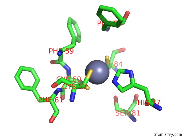
Mono view
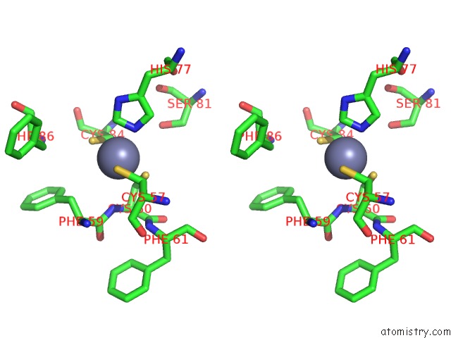
Stereo pair view
Mono view
Stereo pair view
A full contact list of Zinc with other atoms in the Zn binding
site number 1 of Crystal Structure of Human Survivin Mutant K62Y/H80W in Complex with H3(1-10) Peptide within 5.0Å range:
|
Zinc binding site 2 out of 2 in 3uik
Go back to
Zinc binding site 2 out
of 2 in the Crystal Structure of Human Survivin Mutant K62Y/H80W in Complex with H3(1-10) Peptide
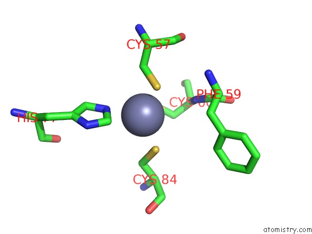
Mono view
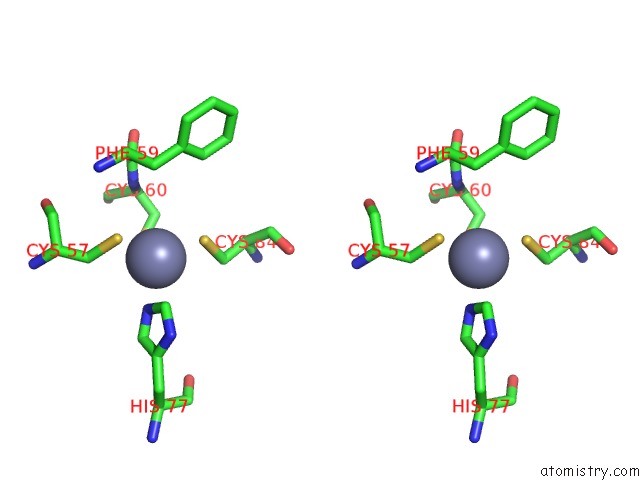
Stereo pair view
Mono view
Stereo pair view
A full contact list of Zinc with other atoms in the Zn binding
site number 2 of Crystal Structure of Human Survivin Mutant K62Y/H80W in Complex with H3(1-10) Peptide within 5.0Å range:
|
Reference:
J.Du,
A.E.Kelly,
H.Funabiki,
D.J.Patel.
Structural Basis For Recognition of H3T3PH and Smac/Diablo N-Terminal Peptides By Human Survivin. Structure V. 20 185 2012.
ISSN: ISSN 0969-2126
PubMed: 22244766
DOI: 10.1016/J.STR.2011.12.001
Page generated: Sat Oct 26 17:22:28 2024
ISSN: ISSN 0969-2126
PubMed: 22244766
DOI: 10.1016/J.STR.2011.12.001
Last articles
Zn in 9JYWZn in 9IR4
Zn in 9IR3
Zn in 9GMX
Zn in 9GMW
Zn in 9JEJ
Zn in 9ERF
Zn in 9ERE
Zn in 9EGV
Zn in 9EGW