Zinc »
PDB 3t65-3ten »
3t9h »
Zinc in PDB 3t9h: Kainate Bound to A Double Cysteine Mutant (A452C/S652C) of the Ligand Binding Domain of GLUA2
Protein crystallography data
The structure of Kainate Bound to A Double Cysteine Mutant (A452C/S652C) of the Ligand Binding Domain of GLUA2, PDB code: 3t9h
was solved by
A.H.Ahmed,
S.Wang,
H.H.Chuang,
R.E.Oswald,
with X-Ray Crystallography technique. A brief refinement statistics is given in the table below:
| Resolution Low / High (Å) | 41.37 / 2.02 |
| Space group | P 2 21 21 |
| Cell size a, b, c (Å), α, β, γ (°) | 47.841, 113.918, 164.786, 90.00, 90.00, 90.00 |
| R / Rfree (%) | 18.7 / 23.5 |
Zinc Binding Sites:
The binding sites of Zinc atom in the Kainate Bound to A Double Cysteine Mutant (A452C/S652C) of the Ligand Binding Domain of GLUA2
(pdb code 3t9h). This binding sites where shown within
5.0 Angstroms radius around Zinc atom.
In total 5 binding sites of Zinc where determined in the Kainate Bound to A Double Cysteine Mutant (A452C/S652C) of the Ligand Binding Domain of GLUA2, PDB code: 3t9h:
Jump to Zinc binding site number: 1; 2; 3; 4; 5;
In total 5 binding sites of Zinc where determined in the Kainate Bound to A Double Cysteine Mutant (A452C/S652C) of the Ligand Binding Domain of GLUA2, PDB code: 3t9h:
Jump to Zinc binding site number: 1; 2; 3; 4; 5;
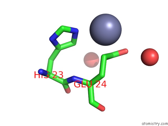
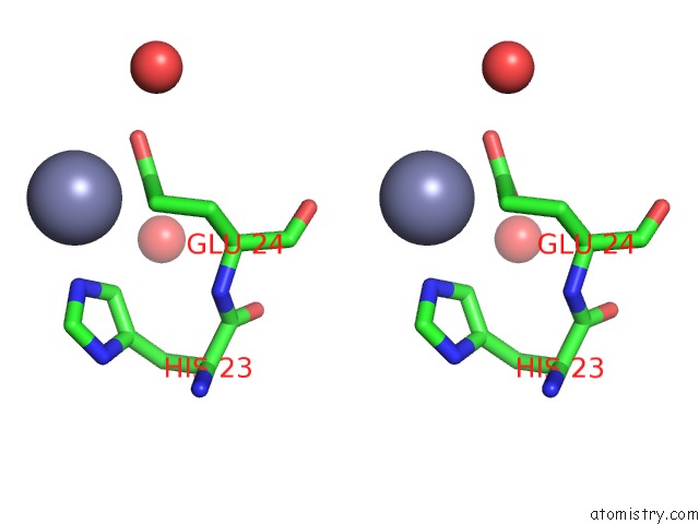
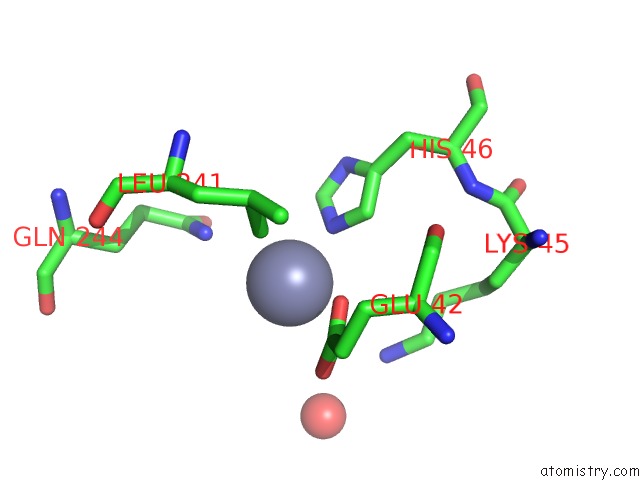
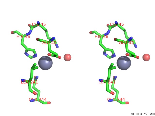
Zinc binding site 1 out of 5 in 3t9h
Go back to
Zinc binding site 1 out
of 5 in the Kainate Bound to A Double Cysteine Mutant (A452C/S652C) of the Ligand Binding Domain of GLUA2
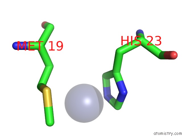
Mono view
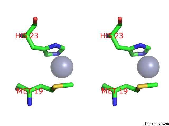
Stereo pair view
Mono view
Stereo pair view
A full contact list of Zinc with other atoms in the Zn binding
site number 1 of Kainate Bound to A Double Cysteine Mutant (A452C/S652C) of the Ligand Binding Domain of GLUA2 within 5.0Å range:
|
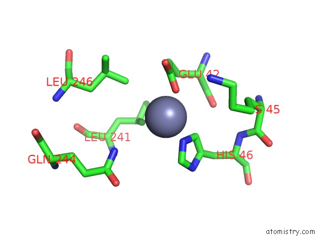
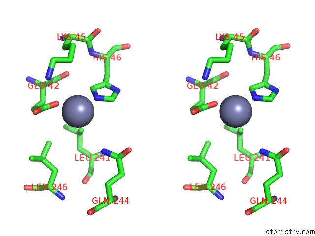
Zinc binding site 2 out of 5 in 3t9h
Go back to
Zinc binding site 2 out
of 5 in the Kainate Bound to A Double Cysteine Mutant (A452C/S652C) of the Ligand Binding Domain of GLUA2
Mono view
Stereo pair view
Mono view
Stereo pair view
A full contact list of Zinc with other atoms in the Zn binding
site number 2 of Kainate Bound to A Double Cysteine Mutant (A452C/S652C) of the Ligand Binding Domain of GLUA2 within 5.0Å range:
|
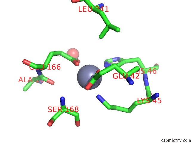
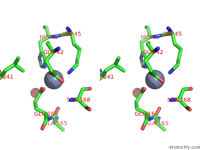
Zinc binding site 3 out of 5 in 3t9h
Go back to
Zinc binding site 3 out
of 5 in the Kainate Bound to A Double Cysteine Mutant (A452C/S652C) of the Ligand Binding Domain of GLUA2
Mono view
Stereo pair view
Mono view
Stereo pair view
A full contact list of Zinc with other atoms in the Zn binding
site number 3 of Kainate Bound to A Double Cysteine Mutant (A452C/S652C) of the Ligand Binding Domain of GLUA2 within 5.0Å range:
|
Zinc binding site 4 out of 5 in 3t9h
Go back to
Zinc binding site 4 out
of 5 in the Kainate Bound to A Double Cysteine Mutant (A452C/S652C) of the Ligand Binding Domain of GLUA2
Mono view
Stereo pair view
Mono view
Stereo pair view
A full contact list of Zinc with other atoms in the Zn binding
site number 4 of Kainate Bound to A Double Cysteine Mutant (A452C/S652C) of the Ligand Binding Domain of GLUA2 within 5.0Å range:
|
Zinc binding site 5 out of 5 in 3t9h
Go back to
Zinc binding site 5 out
of 5 in the Kainate Bound to A Double Cysteine Mutant (A452C/S652C) of the Ligand Binding Domain of GLUA2
Mono view
Stereo pair view
Mono view
Stereo pair view
A full contact list of Zinc with other atoms in the Zn binding
site number 5 of Kainate Bound to A Double Cysteine Mutant (A452C/S652C) of the Ligand Binding Domain of GLUA2 within 5.0Å range:
|
Reference:
A.H.Ahmed,
S.Wang,
H.H.Chuang,
R.E.Oswald.
Mechanism of Ampa Receptor Activation By Partial Agonists: Disulfide Trapping of Closed Lobe Conformations. J.Biol.Chem. V. 286 35257 2011.
ISSN: ISSN 0021-9258
PubMed: 21846932
DOI: 10.1074/JBC.M111.269001
Page generated: Sat Oct 26 16:24:25 2024
ISSN: ISSN 0021-9258
PubMed: 21846932
DOI: 10.1074/JBC.M111.269001
Last articles
Zn in 9JYWZn in 9IR4
Zn in 9IR3
Zn in 9GMX
Zn in 9GMW
Zn in 9JEJ
Zn in 9ERF
Zn in 9ERE
Zn in 9EGV
Zn in 9EGW