Zinc »
PDB 3qm3-3qz6 »
3qw6 »
Zinc in PDB 3qw6: Crystal Structure of the Protease Domain of Botulinum Neurotoxin Serotype A with A Peptide Inhibitor Rygc
Enzymatic activity of Crystal Structure of the Protease Domain of Botulinum Neurotoxin Serotype A with A Peptide Inhibitor Rygc
All present enzymatic activity of Crystal Structure of the Protease Domain of Botulinum Neurotoxin Serotype A with A Peptide Inhibitor Rygc:
3.4.24.69;
3.4.24.69;
Protein crystallography data
The structure of Crystal Structure of the Protease Domain of Botulinum Neurotoxin Serotype A with A Peptide Inhibitor Rygc, PDB code: 3qw6
was solved by
D.Kumaran,
S.Swaminathan,
with X-Ray Crystallography technique. A brief refinement statistics is given in the table below:
| Resolution Low / High (Å) | 35.99 / 1.60 |
| Space group | P 1 21 1 |
| Cell size a, b, c (Å), α, β, γ (°) | 51.066, 66.376, 64.805, 90.00, 98.38, 90.00 |
| R / Rfree (%) | 20.5 / 22.2 |
Other elements in 3qw6:
The structure of Crystal Structure of the Protease Domain of Botulinum Neurotoxin Serotype A with A Peptide Inhibitor Rygc also contains other interesting chemical elements:
| Sodium | (Na) | 1 atom |
Zinc Binding Sites:
The binding sites of Zinc atom in the Crystal Structure of the Protease Domain of Botulinum Neurotoxin Serotype A with A Peptide Inhibitor Rygc
(pdb code 3qw6). This binding sites where shown within
5.0 Angstroms radius around Zinc atom.
In total only one binding site of Zinc was determined in the Crystal Structure of the Protease Domain of Botulinum Neurotoxin Serotype A with A Peptide Inhibitor Rygc, PDB code: 3qw6:
In total only one binding site of Zinc was determined in the Crystal Structure of the Protease Domain of Botulinum Neurotoxin Serotype A with A Peptide Inhibitor Rygc, PDB code: 3qw6:
Zinc binding site 1 out of 1 in 3qw6
Go back to
Zinc binding site 1 out
of 1 in the Crystal Structure of the Protease Domain of Botulinum Neurotoxin Serotype A with A Peptide Inhibitor Rygc
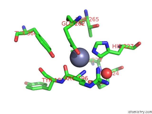
Mono view
Stereo pair view
Mono view
Stereo pair view
A full contact list of Zinc with other atoms in the Zn binding
site number 1 of Crystal Structure of the Protease Domain of Botulinum Neurotoxin Serotype A with A Peptide Inhibitor Rygc within 5.0Å range:
|
Reference:
G.Kumar,
D.Kumaran,
S.A.Ahmed,
S.Swaminathan.
Peptide Inhibitors of Botulinum Neurotoxin Serotype A: Design, Inhibition, Cocrystal Structures, Structure-Activity Relationship and Pharmacophore Modeling. Acta Crystallogr.,Sect.D V. 68 511 2012.
ISSN: ISSN 0907-4449
PubMed: 22525749
DOI: 10.1107/S0907444912003551
Page generated: Sat Oct 26 12:23:13 2024
ISSN: ISSN 0907-4449
PubMed: 22525749
DOI: 10.1107/S0907444912003551
Last articles
Zn in 9JYWZn in 9IR4
Zn in 9IR3
Zn in 9GMX
Zn in 9GMW
Zn in 9JEJ
Zn in 9ERF
Zn in 9ERE
Zn in 9EGV
Zn in 9EGW