Zinc »
PDB 3qm3-3qz6 »
3qnf »
Zinc in PDB 3qnf: Crystal Structure of the Open State of Human Endoplasmic Reticulum Aminopeptidase 1 ERAP1
Protein crystallography data
The structure of Crystal Structure of the Open State of Human Endoplasmic Reticulum Aminopeptidase 1 ERAP1, PDB code: 3qnf
was solved by
M.Vollmar,
G.Kochan,
T.Krojer,
D.Harvey,
A.Chaikuad,
C.Allerston,
J.R.C.Muniz,
J.Raynor,
E.Ugochukwu,
G.Berridge,
B.P.Wordsworth,
F.Vondelft,
C.Bountra,
C.H.Arrowsmith,
A.Edwards,
K.Kavanagh,
U.Oppermann,
Structural Genomics Consortium (Sgc),
with X-Ray Crystallography technique. A brief refinement statistics is given in the table below:
| Resolution Low / High (Å) | 19.99 / 3.00 |
| Space group | P 21 21 21 |
| Cell size a, b, c (Å), α, β, γ (°) | 97.246, 132.816, 233.687, 90.00, 90.00, 90.00 |
| R / Rfree (%) | 22.9 / 28.3 |
Zinc Binding Sites:
The binding sites of Zinc atom in the Crystal Structure of the Open State of Human Endoplasmic Reticulum Aminopeptidase 1 ERAP1
(pdb code 3qnf). This binding sites where shown within
5.0 Angstroms radius around Zinc atom.
In total 3 binding sites of Zinc where determined in the Crystal Structure of the Open State of Human Endoplasmic Reticulum Aminopeptidase 1 ERAP1, PDB code: 3qnf:
Jump to Zinc binding site number: 1; 2; 3;
In total 3 binding sites of Zinc where determined in the Crystal Structure of the Open State of Human Endoplasmic Reticulum Aminopeptidase 1 ERAP1, PDB code: 3qnf:
Jump to Zinc binding site number: 1; 2; 3;
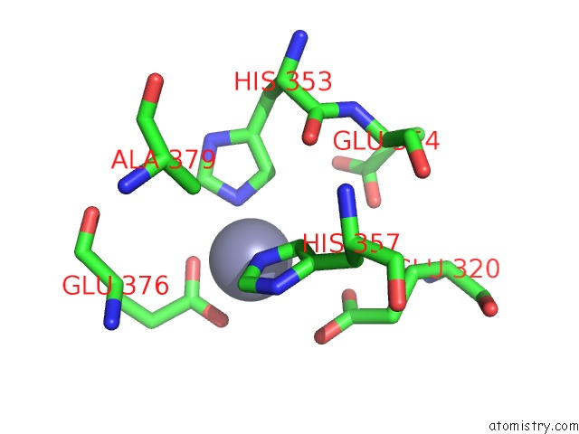
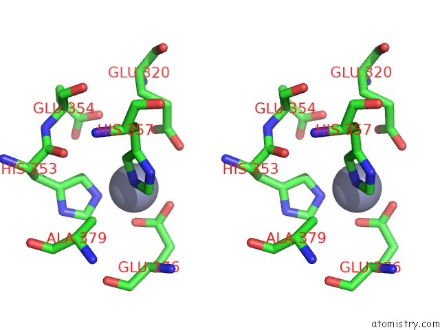
Zinc binding site 1 out of 3 in 3qnf
Go back to
Zinc binding site 1 out
of 3 in the Crystal Structure of the Open State of Human Endoplasmic Reticulum Aminopeptidase 1 ERAP1
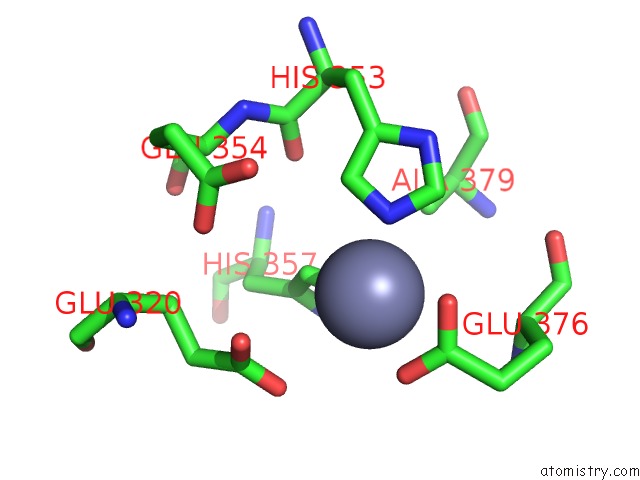
Mono view
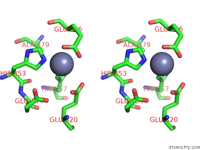
Stereo pair view
Mono view
Stereo pair view
A full contact list of Zinc with other atoms in the Zn binding
site number 1 of Crystal Structure of the Open State of Human Endoplasmic Reticulum Aminopeptidase 1 ERAP1 within 5.0Å range:
|
Zinc binding site 2 out of 3 in 3qnf
Go back to
Zinc binding site 2 out
of 3 in the Crystal Structure of the Open State of Human Endoplasmic Reticulum Aminopeptidase 1 ERAP1
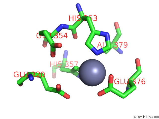
Mono view
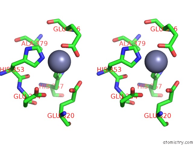
Stereo pair view
Mono view
Stereo pair view
A full contact list of Zinc with other atoms in the Zn binding
site number 2 of Crystal Structure of the Open State of Human Endoplasmic Reticulum Aminopeptidase 1 ERAP1 within 5.0Å range:
|
Zinc binding site 3 out of 3 in 3qnf
Go back to
Zinc binding site 3 out
of 3 in the Crystal Structure of the Open State of Human Endoplasmic Reticulum Aminopeptidase 1 ERAP1
Mono view
Stereo pair view
Mono view
Stereo pair view
A full contact list of Zinc with other atoms in the Zn binding
site number 3 of Crystal Structure of the Open State of Human Endoplasmic Reticulum Aminopeptidase 1 ERAP1 within 5.0Å range:
|
Reference:
G.Kochan,
T.Krojer,
D.Harvey,
R.Fischer,
L.Chen,
M.Vollmar,
F.Von Delft,
K.L.Kavanagh,
M.A.Brown,
P.Bowness,
P.Wordsworth,
B.M.Kessler,
U.Oppermann.
Crystal Structures of the Endoplasmic Reticulum Aminopeptidase-1 (ERAP1) Reveal the Molecular Basis For N-Terminal Peptide Trimming. Proc.Natl.Acad.Sci.Usa V. 108 7745 2011.
ISSN: ISSN 0027-8424
PubMed: 21508329
DOI: 10.1073/PNAS.1101262108
Page generated: Sat Oct 26 12:14:34 2024
ISSN: ISSN 0027-8424
PubMed: 21508329
DOI: 10.1073/PNAS.1101262108
Last articles
Zn in 9JYWZn in 9IR4
Zn in 9IR3
Zn in 9GMX
Zn in 9GMW
Zn in 9JEJ
Zn in 9ERF
Zn in 9ERE
Zn in 9EGV
Zn in 9EGW